Eigengrau
Eigengrau (German for "intrinsic gray"; pronounced [ˈʔaɪ̯gŋ̍ˌgʁaʊ̯] (![]() listen)), also called Eigenlicht (Dutch and German for "intrinsic light"), dark light, or brain gray, is the uniform dark gray background color that many people report seeing in the absence of light. The term Eigenlicht dates back to the nineteenth century,[1] and has rarely been used in recent scientific publications. Common scientific terms for the phenomenon include "visual noise" or "background adaptation". These terms arise due to the perception of an ever-changing field of tiny black and white dots seen in the phenomenon.[2]
listen)), also called Eigenlicht (Dutch and German for "intrinsic light"), dark light, or brain gray, is the uniform dark gray background color that many people report seeing in the absence of light. The term Eigenlicht dates back to the nineteenth century,[1] and has rarely been used in recent scientific publications. Common scientific terms for the phenomenon include "visual noise" or "background adaptation". These terms arise due to the perception of an ever-changing field of tiny black and white dots seen in the phenomenon.[2]
Eigengrau is perceived as lighter than a black object in normal lighting conditions, because contrast is more important to the visual system than absolute brightness.[3] For example, the night sky looks darker than Eigengrau because of the contrast provided by the stars.
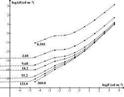
Contrast threshold data, collected by Blackwell[4] and plotted by Crumey, shows Eigengrau occurring at adaptation luminances below approximately 10− 5 cd m−2 (25.08 mag arcsec−2).[5] This is a limiting case of Ricco's law.
Cause
Researchers noticed as early as 1860 that the shape of intensity-sensitivity curves could be explained by assuming that an intrinsic source of noise in the retina produces random events indistinguishable from those triggered by real photons.[6] In human rod cells, these events occur about once every 100 seconds on average, which, taking into account the number of rhodopsin molecules in a rod cell, implies that the half-life of a rhodopsin molecule is about 420 years.[7] The indistinguishability of dark events from photon responses supports this explanation, because rhodopsin is at the input of the transduction chain. On the other hand, processes such as the spontaneous release of neurotransmitters cannot be completely ruled out.[8]
See also
References
- ↑ Ladd, Trumbull (1894). "Direct control of the retinal field.". Psychological Review 1 (4): 351–55. doi:10.1037/h0068980. https://zenodo.org/record/1429102.
- ↑ Hansen RM, Fulton AB (January 2000). "Background adaptation in children with a history of mild retinopathy of prematurity". Invest. Ophthalmol. Vis. Sci. 41 (1): 320–24. PMID 10634637.
- ↑ Wallach, Hans (1948). "Brightness Constancy and the Nature of Achromatic Colors". Journal of Experimental Psychology 38 (3): 310–24. doi:10.1037/h0053804. PMID 18865234.
- ↑ "H. Richard Blackwell, Contrast Thresholds of the Human Eye. Journal of the Optical Society of America Vol. 36, Issue 11, pp. 624-643 (1946)". https://doi.org/10.1364/JOSA.36.000624.
- ↑ Crumey, A. (2014). Human contrast threshold and astronomical visibility. MNRAS 442, 2600–2619.
- ↑ Barlow, H.B. (1980). Two components of electrical dark noise in toad retinal rod outer segments. 309. New York: Springer-Verlag. 591–621. doi:10.1113/jphysiol.1980.sp013529. ISBN 978-0-387-05146-8.
- ↑ Baylor, Denis A. (1 January 1987). "Photoreceptor Signals and Vision". Investigative Ophthalmology & Visual Science 28 (1): 34–49. PMID 3026986. http://www.iovs.org/cgi/reprint/28/1/34.
- ↑ Shapley, Robert; Enroth-Cugell, Christina (1984). "Visual Adaptation and Retinal Gain Controls". Progress in Retinal Research 3: 263–346. doi:10.1016/0278-4327(84)90011-7.
Color topics | ||||||||
|---|---|---|---|---|---|---|---|---|
| Color science |
|  | ||||||
| Color philosophy |
| |||||||
| Color terms |
| |||||||
| Color organizations | ||||||||
| Lists | ||||||||
| Related | ||||||||
 |  |

