File:Pyxidiophora.jpg

From HandWiki
Original file(1,138 × 1,900 pixels, file size: 563 KB, MIME type: image/jpeg)

This file is from a shared repository and may be used by other projects. The description on its file description page there is shown below.

Summary

Description
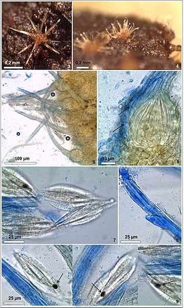
English: Pyxidiophora arvernensis. 3–5. Perithecia on dung. 6. A perithecium under optical microscope; arrows point to the exoperidial wall.

7. Ascospores, the arrow points to the acute apex. 8. The ascospore stained with cotton blue. 9. Adhesive discs present in the region near the apex of

mature ascospores (arrows)
Português: Pyxidiophora arvernensis. 3–5. Perithecia on dung. 6. A perithecium under optical microscope; arrows point to the exoperidial wall.

7. Ascospores, the arrow points to the acute apex. 8. The ascospore stained with cotton blue. 9. Adhesive discs present in the region near the apex of

mature ascospores (arrows)
Source Own work
Author Franciscocalaça

Licensing

I, the copyright holder of this work, hereby publish it under the following license:
w:en:Creative Commons
attribution
This file is licensed under the Creative Commons Attribution 4.0 International license.
You are free:
  • to share – to copy, distribute and transmit the work
  • to remix – to adapt the work
Under the following conditions:
  • attribution – You must give appropriate credit, provide a link to the license, and indicate if changes were made. You may do so in any reasonable manner, but not in any way that suggests the licensor endorses you or your use.

Captions

Add a one-line explanation of what this file represents

File history

Click on a date/time to view the file as it appeared at that time.

Date/TimeThumbnailDimensionsUserComment
current14:40, 23 September 2017Thumbnail for version as of 14:40, 23 September 20171,138 × 1,900 (563 KB)imagescommonswiki>Franciscocalaça{{Information |Description ={{en|1=Pyxidiophora arvernensis. 3–5. Perithecia on dung. 6. A perithecium under optical microscope; arrows point to the exoperidial wall. 7. Ascospores, the arrow points to the acute apex. 8. The ascospore stained with...

The following file is a duplicate of this file (more details):

The following 2 pages use this file: