File:Triangle of Koch (large).jpg
From HandWiki

Size of this preview: 671 × 599 pixels. Other resolutions: 537 × 480 pixels | 1,594 × 1,424 pixels.
Original file (1,594 × 1,424 pixels, file size: 220 KB, MIME type: image/jpeg)
Summary
| Description |
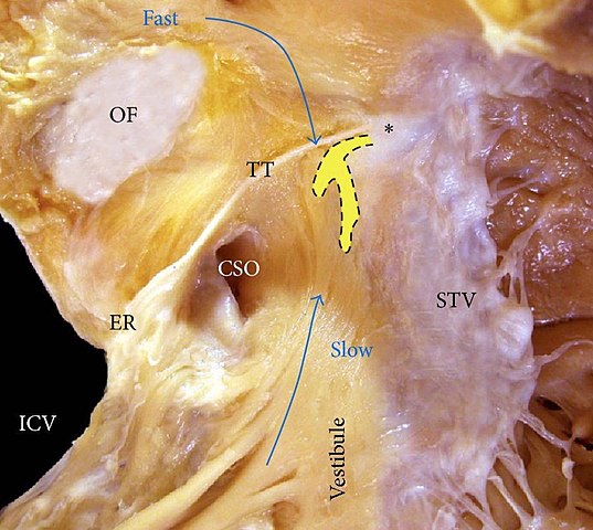
English: Dissection in right anterior oblique view of the right atrium shows the borders of the triangle of Koch. In this view, the putative fast and slow pathways toward the AV node (dotted shape in yellow) are depicted. Asterisk (*): central fibrous body, CSO: coronary sinus ostium, ER: Eustachian ridge, ICV: inferior cava vein, OF: oval fossa, STV: septal leaflet of the tricuspid valve, and TT: tendon of Todaro. Deutsch: Dissektion in rechter anteriorer Schrägansicht des rechten Vorhofs mit den Grenzen des Koch-Dreiecks. In dieser Ansicht sind die mutmaßlichen schnellen und langsamen Bahnen zum AV-Knoten (gestrichelt in gelb) dargestellt. Sternchen (*): zentraler Faserkörper, CSO: Einmündung des Sinus coronarius (Ostium sinus coronarii), ER: Valvula Eustachii (Valvula venae cavae inferioris), ICV: Vena cava inferior, OF: Fossa ovalis, STV: kammerscheidewandseitiges Tricuspidalis-Segel und TT: Todarosehne. |
| Date | |
| Source | Damián Sánchez-Quintana, Manuel Doblado-Calatrava, José Angel Cabrera, Yolanda Macías, Farhood Saremi: Anatomical Basis for the Cardiac Interventional Electrophysiologist. In: BioMed Research International. 2015, Band 2015, S. 1–24 doi:10.1155/2015/547364. |
| Author | Damián Sánchez-Quintana, Manuel Doblado-Calatrava, José Angel Cabrera, Yolanda Macías, Farhood Saremi |
| Permission (Reusing this file) |
open access publication: https://www.hindawi.com/journals/bmri/2015/547364/#copyright |
Licensing
This file is licensed under the Creative Commons Attribution 3.0 Unported license.
- You are free:
- to share – to copy, distribute and transmit the work
- to remix – to adapt the work
- Under the following conditions:
- attribution – You must give appropriate credit, provide a link to the license, and indicate if changes were made. You may do so in any reasonable manner, but not in any way that suggests the licensor endorses you or your use.
File history
Click on a date/time to view the file as it appeared at that time.
| Date/Time | Thumbnail | Dimensions | User | Comment | |
|---|---|---|---|---|---|
| current | 18:41, 3 December 2022 | | 1,594 × 1,424 (220 KB) | imagescommonswiki>Polarlys | {{Information |Description= |Source=Damián Sánchez-Quintana, Manuel Doblado-Calatrava, José Angel Cabrera, Yolanda Macías, Farhood Saremi: ''Anatomical Basis for the Cardiac Interventional Electrophysiologist.'' In: ''BioMed Research International.'' 2015, Band 2015, S. 1–24 {{DOI|10.1155/2015/547364}}. |Date=2015 |Author=Damián Sánchez-Quintana, Manuel Doblado-Calatrava, José Angel Cabrera, Yolanda Macías, Farhood Saremi |Permission=open access publication: https://www.hindawi.com/journ... |
File usage
The following file is a duplicate of this file (more details):
- File:Triangle of Koch (large).jpg from Wikimedia Commons
The following page uses this file:


.jpg)
.jpg&action=edit&redlink=1)
.jpg)
.jpg&action=mzwi)
.jpg)
.jpg)
.jpg)
.jpg&action=info)