Biology:Omasum
The omasum, also known as the bible,[1] the fardel,[1] the manyplies[1] and the psalterium,[1] is the third compartment of the stomach in ruminants. The omasum comes after the rumen and reticulum and before the abomasum. Different ruminants have different omasum structures and function based on the food that they eat and how they developed through evolution.[2]
Anatomy
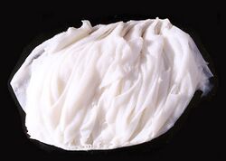
The omasum can be found on the right side of the cranial portion of the rumen.[3] The omasum receives food from the reticulum through the reticulo-omasal orifice[3] and provides food to the abomasum through the omaso-abomasal orifice.[4] The omasum is spherical[5] to crescent shape[6] and has multiple leaflets similar to that of a book[7] called omasal laminae.[4] The omasal laminae are made of thin muscular layers covered with a nonglandular mucous membrane.[4] The omasal laminae come from the sides of the large curvature and project towards the inside of the omasum, extending from the reticulo-omasal orifice to the omaso-abomasal orifice.[8][4] The laminae greatly increase the surface area of the omasum.[3][9] The laminae are covered in omasal papillae that are claw-like in some ruminants or blunt cones in others.[4][2] These papillae further increase the surface area but they also provide increased friction against the food particles.[3]
Function
The function of the omasum is not completely understood.[5] During the second contraction phase of the reticulum, the reticule-omasal sphincter opens for a few seconds allowing a small volume of finely dispersed and well-fermented ingesta to enter the omasum.[3]
The omasum has two physiological compartments: omasal canal that transfers food from the reticulum to the omasum, and the inter-laminate recesses between the mucosal laminae which provide the area for absorption.[2] The omasum is where food particles that are small enough get transferred into the abomasum for enzymatic digestion.[5][2] In ruminants with a more sophisticated omasum, the large surface area[9] allows it to play a key role in the absorption of water, electrolytes,[2][4] volatile fatty acids, minerals, and the fermentation of food.[5]
Young ruminants that are still drinking milk have an esophageal groove that allows milk to bypass the rumen and go straight from the esophagus to the omasum.[10]
Species differences
An early version of the omasum is seen in early ruminants like duikers and muntjacs, where it is a little more than a strainer sieve which prevents un-chewed foods from entering the abomasum.[2]
The smallest omasum belongs to ruminants that consume high quality diets like the moose and roe deer, while the largest belongs to those who are un-selective grass and roughage eaters like cattle and sheep.[2]
The omasum is not only bigger in grass and roughage eaters but there is greater differentiation in the book-like structure; seen as an increase in the number of laminae.[2]
Culinary uses
See also
References
- ↑ 1.0 1.1 1.2 1.3 The Chambers Dictionary, Ninth Edition, Chambers Harrap Publishers, 2003
- ↑ 2.0 2.1 2.2 2.3 2.4 2.5 2.6 2.7 Hofmann, R. (1989). "Evolutionary steps of ecophysiological adaptation and diversification of ruminants: a comparative view of their digestive system". Oecologia 78 (4): 443–457. doi:10.1007/BF00378733. PMID 28312172. Bibcode: 1989Oecol..78..443H. https://www.uky.edu/Ag/AnimalSciences/instruction/asc684/PDF/oecologia78_443.pdf.
- ↑ 3.0 3.1 3.2 3.3 3.4 Sjaastad, Oystein (2010). Physiology of domestic animals. Hove, Knut., Sand, Olav. (2nd ed.). Oslo: Scandinavian Veterinary Press. pp. 555–556. ISBN 978-82-91743-07-3. OCLC 670546738.
- ↑ 4.0 4.1 4.2 4.3 4.4 4.5 Yamamoto, Y.; Kitamura, N. (1994). "Morphological study of the surface structure of the omasal laminae in cattle, sheep and goats". Anatomia, Histologia, Embryologia 23 (2): 166–167. doi:10.1111/j.1439-0264.1994.tb00249.x. ISSN 0340-2096. PMID 7978351.
- ↑ 5.0 5.1 5.2 5.3 Grünberg, Walter; Constable, Peter D. (2009). "Function and Dysfunction of the Ruminant Forestomach". Food Animal Practice (5 ed.). pp. 12–19. doi:10.1016/b978-141603591-6.10006-5. ISBN 9781416035916.
- ↑ Braun, Ueli (2009). "Ultrasonography of the Gastrointestinal Tract in Cattle". Veterinary Clinics of North America: Food Animal Practice 25 (3): 567–590. doi:10.1016/j.cvfa.2009.07.004. PMID 19825434. https://www.zora.uzh.ch/id/eprint/26375/1/Braun.pdf.
- ↑ Stumpff, Friederike; Georgi, Maria-Ifigenia; Mundhenk, Lars; Rabbani, Imtiaz; Fromm, Michael; Martens, Holger; Günzel, Dorothee (2011-09-01). "Sheep rumen and omasum primary cultures and source epithelia: barrier function aligns with expression of tight junction proteins" (in en). Journal of Experimental Biology 214 (17): 2871–2882. doi:10.1242/jeb.055582. ISSN 0022-0949. PMID 21832130.
- ↑ Green, E.; Baker, C. (1996). "The surface morphology of the omasum of the African goat". Journal of the South African Veterinary Association 67 (3): 117–122. ISSN 0038-2809. PMID 9120853.
- ↑ 9.0 9.1 "Animal Structure & Function". http://sci.waikato.ac.nz/farm/content/animalstructure.html.
- ↑ "Rumen Physiology and Rumination" (in en). http://www.vivo.colostate.edu/hbooks/pathphys/digestion/herbivores/rumination.html.
 |


