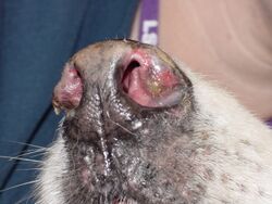
Medicine:Canine discoid lupus erythematosus
Discoid lupus erythematosus (DLE) is an uncommon autoimmune disease of the basal cell layer of the skin. It occurs in humans[1] and cats, more frequently occurring in dogs. It was first described in dogs by Griffin and colleagues in 1979.[2][3] DLE is one form of cutaneous lupus erythematosus (CLE). DLE occurs in dogs in two forms: a classical facial predominant form or generalized with other areas of the body affected. Other non-discoid variants of CLE include vesicular CLE, exfoliative CLE and mucocutaneous CLE.[4] It does not progress to systemic lupus erythematosus (SLE) in dogs. SLE can also have skin symptoms, but it appears that the two are either separate diseases.[5] DLE in dogs differs from SLE in humans in that plasma cells predominate histologically instead of T lymphocytes.[6] Because worsening of symptoms occurs with increased ultraviolet light exposure, sun exposure most likely plays a role in DLE, although certain breeds (see below) are predisposed.[7] After pemphigus foliaceus, DLE is the second most common autoimmune skin disease in dogs.[8]
Symptoms
The most common initial symptom is scaling and loss of pigment on the nose. The surface of the nose becomes smooth gray, and ulcerated, instead of the normal black cobblestone texture. Over time the lips, the skin around the eyes, the ears, and the genitals may become involved.[9] Lesions may progress to ulceration and lead to tissue destruction. DLE is often worse in summer due to increased sun exposure.
Diagnosis
DLE is easily confused with solar dermatitis, pemphigus, ringworm, and other types of dermatitis. Biopsy is required to make the distinction. Histopathologically, there is inflammation at the dermoepidermal junction and degeneration of the basal cell layer.[8] Unlike in SLE, an anti-nuclear antibody test is usually negative.[5]
Treatment
Avoiding sun exposure and the use of sunscreens (not containing zinc oxide as this is toxic to dogs[10][11]) is important. Topical therapy includes corticosteroid and tacrolimus[12] use. Oral vitamin E or omega-3 and omega-6 fatty acids are also used. More refractory cases may require the use of oral niacinamide and tetracycline or immuno-suppressive medication such as corticosteroids, azathioprine, or chlorambucil.[13] Treatment is often lifelong, but there is a good prognosis for long-term remission.
Commonly affected dog breed
- Alaskan Malamute
- Collie
- German Shepherd Dog
- Shetland Sheepdog
- Siberian Husky
- Brittany
- German Shorthaired Pointer[6]
- Africanis
- Rottweilers
References
- ↑ American Osteopathic College of Dermatology, "Discoid Lupus Erythematosus".
- ↑ Griffin, C.E.; Stannard, A.A.; Ihrke, P.J.; Ardans, A.A.; Cello, R.M.; Bjorling, D.R. (November 1979). "Canine discoid lupus erythematosus" (in en). Veterinary Immunology and Immunopathology 1 (1): 79–87. doi:10.1016/0165-2427(79)90009-6. PMID 15612271.
- ↑ Olivry, Thierry; Linder, Keith E.; Banovic, Frane (December 2018). "Cutaneous lupus erythematosus in dogs: a comprehensive review" (in en). BMC Veterinary Research 14 (1): 132. doi:10.1186/s12917-018-1446-8. ISSN 1746-6148. PMID 29669547.
- ↑ Banovic, Frane (January 2019). "Canine Cutaneous Lupus Erythematosus" (in en). Veterinary Clinics of North America: Small Animal Practice 49 (1): 37–45. doi:10.1016/j.cvsm.2018.08.004. PMID 30227971.
- ↑ 5.0 5.1 Ettinger, Stephen J.; Feldman, Edward C. (1995). Textbook of Veterinary Internal Medicine (4th ed.). W.B. Saunders Company. ISBN 0-7216-6795-3. OCLC 28723106.
- ↑ 6.0 6.1 Griffin, Craig E.; Miller, William H.; Scott, Danny W. (2001). Small Animal Dermatology (6th ed.). W.B. Saunders Company. ISBN 0-7216-7618-9. OCLC 43845516.
- ↑ Mueller, Ralf S. (2005). "Immune-mediated skin diseases" (PDF). Proceedings of the 50° Congresso Nazionale Multisala SCIVAC. http://www.ivis.org/proceedings/scivac/2005/Mueller1_en.pdf?LA=1. Retrieved 2007-03-13.
- ↑ 8.0 8.1 Osborn, S. (2006). "Autoimmune Diseases in the Dog". Proceedings of the North American Veterinary Conference. http://www.ivis.org/proceedings/NAVC/2006/SAE/128.asp?LA=1. Retrieved 2007-03-13.
- ↑ "A case of interface perianal dermatitis in a dog: is this an unusual manifestation of lupus erythematosus?". Vet Pathol 43 (5): 761–4. 2006. doi:10.1354/vp.43-5-761. PMID 16966456.
- ↑ Kiriluk, Ellaine (2019-07-17). "Protect your dog from the extreme heat of summer" (in en-US). Daily Herald (Arlington Heights, Illinois). https://www.dailyherald.com/submitted/20190717/protect-your-dog-from-the-extreme-heat-of-summer. "Human sunscreen is safe if it doesn't contain zinc oxide, which can be toxic if ingested. Since there are ingredients in sunscreens that can be toxic to both cats and dogs, [Dr. Ruth MacPete, DVM] recommends using only a veterinarian-approved sunscreen."
- ↑ Devera, Carina (2019-07-08). "Beware the hidden toxins that seem harmless but can be deadly" (in en-US). Marin Independent Journal (San Rafael, California). https://www.marinij.com/2019/07/08/beware-the-hidden-toxins-that-seem-harmless-but-can-be-deadly/. "Zinc oxide is effective as a sunscreen for humans, but it’s toxic for dogs. If ingested, it can damage your dog’s red blood cells."
- ↑ Griffies, J. D.; Mendelsohn, C. L.; Rosenkrantz, W. S.; Muse, R.; Boord, M. J.; Griffin, C. E. (August 2002). "Topical 0.1% tacrolimus for the treatment of discoid lupus erythematosus and pemphigus erythematosus in dogs" (in en). Veterinary Dermatology 13 (4): 211–229. doi:10.1046/j.1365-3164.2002.00298_3.x. ISSN 0959-4493.
- ↑ "Topical 0.1% tacrolimus for the treatment of discoid lupus erythematosus and pemphigus erythematosus in dogs". J Am Anim Hosp Assoc 40 (1): 29–41. 2004. doi:10.5326/0400029. PMID 14736903.
 |


