Biology:Stellate cell
| Stellate cell | |
|---|---|
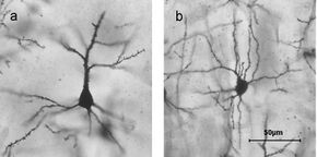
 Golgi stained cortical neurons A) Layer II/III pyramidal cell B) layer IV spiny stellate cell | |
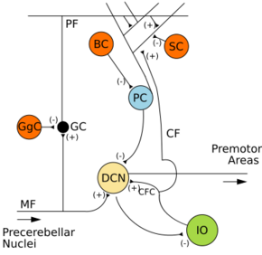
 Microcircuitry of the cerebellum. Excitatory synapses are denoted by (+) and inhibitory synapses by (-). MF: Mossy fiber. DCN: Deep cerebellar nuclei. IO: Inferior olive. CF: Climbing fiber. GC: Granule cell. PF: Parallel fiber. PC: Purkinje cell. GgC: Golgi cell. SC: Stellate cell. BC: Basket cell. | |
| Anatomical terms of neuroanatomy |
Stellate cells are neurons in the central nervous system, named for their star-like shape formed by dendritic processes radiating from the cell body. Many stellate cells are GABAergic and are located in the molecular layer of the cerebellum.[1] Stellate cells are derived from dividing progenitor cells in the white matter of postnatal cerebellum. Dendritic trees can vary between neurons. There are two types of dendritic trees in the cerebral cortex, which include pyramidal cells, which are pyramid shaped and stellate cells which are star shaped. Dendrites can also aid neuron classification. Dendrites with spines are classified as spiny, those without spines are classified as aspinous.[2] Stellate cells can be spiny or aspinous, while pyramidal cells are always spiny. Most common stellate cells are the inhibitory interneurons found within the upper half of the molecular layer in the cerebellum.[3] Cerebellar stellate cells synapse onto the dendritic trees of Purkinje cells and send inhibitory signals.[4] Stellate neurons are sometimes found in other locations in the central nervous system; cortical spiny stellate cells are found in layer IVC of the primary visual cortex.[2] In the somatosensory barrel cortex of mice and rats, glutamatergic (excitatory) spiny stellate cells are organized in the barrels of layer 4.[5] They receive excitatory synaptic fibres from the thalamus and process feed forward excitation to 2/3 layer of the primary visual cortex to pyramidal cells. Cortical spiny stellate cells have a 'regular' firing pattern. Stellate cells are chromophobes, that is cells that does not stain readily, and thus appears relatively pale under the microscope.
Cerebellar stellate cells are inhibitory and GABAergic.[6] Stellate and basket cells originate from the cerebellar ventricular zone (CVZ) along with Purkinje cells and Bergmann glia[7][8] Due to their similarity, basket and stellate cells are grouped together when examined during migration, especially given they follow the same pathway. After mitosis, these cells start in the deep layer of the white matter and migrate up through the internal granular layer (IGL) and purkinje cell layer (PCL)[9] until they reach the molecular layer. During their time in the molecular layer, they change orientation and positioning until they eventually end up in the middle portion of this layer, facing the rostrocaudal direction.[10] Once in this layer, the stellate cells are guided to their correct placement by Bergman glial cells.[11]
GABAergic aspinous stellate cells are found in the somatosensory cortex. Apart from visual classification of the aspinous dendrites, they can be immunohistochemically labelled with glutamic acid decarboxylase(GAD) because of their GABAergic activity, and occasionally colocalize with neuropeptides.[12]
See also
- Stellate ganglion
- List of distinct cell types in the adult human body
References
- ↑ Patterning and Cell Type Specification in the Developing CNS and PNS : Comprehensive Developmental Neuroscience.. Elsevier Science. 2013. p. 215. ISBN 978-0-12-397265-1. https://public.ebookcentral.proquest.com/choice/publicfullrecord.aspx?p=1187144.[yes|permanent dead link|dead link}}]
- ↑ 2.0 2.1 Costa, Nuno Maçarico da; Martin, Kevan A. C. (2011-02-23). "How Thalamus Connects to Spiny Stellate Cells in the Cat's Visual Cortex" (in en). Journal of Neuroscience 31 (8): 2925–2937. doi:10.1523/JNEUROSCI.5961-10.2011. ISSN 0270-6474. PMID 21414914.
- ↑ Patterning and Cell Type Specification in the Developing CNS and PNS : Comprehensive Developmental Neuroscience. (1 ed.). Elsevier Science. 2013. ISBN 978-0-12-397265-1. https://public.ebookcentral.proquest.com/choice/publicfullrecord.aspx?p=1187144.[yes|permanent dead link|dead link}}]
- ↑ Chan-Palay, Victoria; Palay, Sanford L. (1972-01-01). "The stellate cells of the rat's cerebellar cortex" (in en). Zeitschrift für Anatomie und Entwicklungsgeschichte 136 (2): 224–248. doi:10.1007/BF00519180. ISSN 0044-2232. PMID 5042759.
- ↑ Petersen, Carl C.H. (October 2007). "The Functional Organization of the Barrel Cortex". Neuron 56 (2): 339–355. doi:10.1016/j.neuron.2007.09.017. ISSN 0896-6273. PMID 17964250.
- ↑ Rubenstein, John; Rakic, Pasko; Rubenstein, John (2013-05-06). Patterning and cell type specification in the developing CNS and PNS : comprehensive developmental neuroscience (1st ed.). Academic Press. ISBN 9780123973481. https://public.ebookcentral.proquest.com/choice/publicfullrecord.aspx?p=1187144.[yes|permanent dead link|dead link}}]
- ↑ Comprehensive developmental neuroscience. Cellular migration and formation of neuronal connections (First ed.). Elsevier Science & Technology. 2013. pp. 283. ISBN 978-0-12-397266-8. https://public.ebookcentral.proquest.com/choice/publicfullrecord.aspx?p=1187145.
- ↑ "Cerebellar Ventricular Zone - Cellular Development, Function & Anatomy - LifeMap Discovery". LifeMap Sciences. https://discovery.lifemapsc.com/in-vivo-development/neural-tube/cerebellar-ventricular-zone.
- ↑ Comprehensive developmental neuroscience. Cellular migration and formation of neuronal connections (First ed.). Elsevier Science and Technology. 2013. pp. 284. ISBN 978-0-12-397266-8. https://public.ebookcentral.proquest.com/choice/publicfullrecord.aspx?p=1187145.
- ↑ Rubenstein, John (2013-05-06). Patterning and Cell Type Specification in the Developing CNS and PNS : Comprehensive Developmental Neuroscience. Elsevier Science & Technology. ISBN 9780123973481.
- ↑ Rubenstein, John (2013-05-06). Neural Circuit Development and Function in the Brain : Comprehensive Developmental Neuroscience. Elsevier Science & Technology. ISBN 9780123973467.
- ↑ Conn, P. Michael (2008). Neuroscience in medicine (3rd ed.). Beaverton, OR: Humana Press. ISBN 9781603274548. https://public.ebookcentral.proquest.com/choice/publicfullrecord.aspx?p=372379.
External links
 |

