Biology:Walthard cell rest

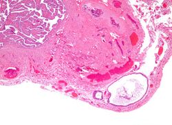
Walthard cell rests, sometimes called Walthard cell nests, are a benign cluster of epithelial cells most commonly found in the connective tissue of the fallopian tubes, but also seen in the mesovarium, mesosalpinx and ovarian hilus.
Appearance

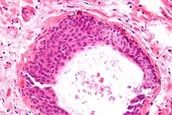
They appear as white/yellow cysts or nodules that can reach a size of 2 millimeters. They typically have elliptical nuclei with a long groove (along the major axis) – so-called "coffee bean" nuclei.
Pathology
It has been suggested that these cell rests are the histogenetic origins of Brenner tumors, due to the histological similarity of the epithelium of Walthard cell rests and Brenner tumors to the urothelium of the lower urinary tract. Also, it has been proposed that Brenner tumors and Walthard cell rests signify urothelial differentiation within the female genital tract.
Eponym
They are named after Swiss gynecologist Max Walthard (1867–1933), who provided a comprehensive description of them in 1903.
Additional images
High magnification micrograph of a Brenner tumor showing the characteristic coffee bean nuclei which are also seen in Walthard cell rests. H&E stain.
References
- Pathology Outline, Fallopian Tubes
- "Immunohistochemical analysis of uroplakins, urothelial specific proteins, in ovarian Brenner tumors, normal tissues, and benign and neoplastic lesions of the female genital tract". Am. J. Pathol. 155 (4): 1047–50. October 1999. doi:10.1016/S0002-9440(10)65206-6. PMID 10514386. PMC 1867018. http://ajp.amjpathol.org/cgi/pmidlookup?view=long&pmid=10514386.
- Wrong Diagnosis.com, Brenner tumors
 |


