Biology:Aleutian disease
| Carnivore amdoparvovirus 1 | |
|---|---|
| Virus classification | |
| (unranked): | Virus |
| Realm: | Monodnaviria |
| Kingdom: | Shotokuvirae |
| Phylum: | Cossaviricota |
| Class: | Quintoviricetes |
| Order: | Piccovirales |
| Family: | Parvoviridae |
| Genus: | Amdoparvovirus |
| Species: | Carnivore amdoparvovirus 1
|
| Synonyms[1] | |
|
Aleutian mink disease virus | |
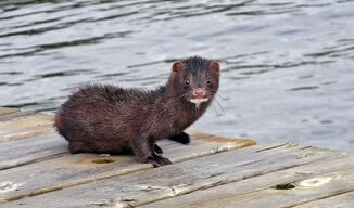
Aleutian disease, also known as mink plasmacytosis, is a disease which causes spontaneous abortion and death in minks and ferrets. It is caused by Carnivore amdoparvovirus 1 (also known as Aleutian disease virus, ADV), a highly contagious parvovirus in the genus Amdoparvovirus. The virus has been found as a natural infection in the Mustelidae family within mink, ferrets, otters, polecats, stone and pine martens and within other carnivores such as skunks, genets, foxes and raccoons.[2][3] This is most commonly explained as because they all share resources and habitats.[3]
History
Aleutian disease was first recognized in ranch-raised mink in 1956. The disease was so named because it was first found in mink with the Aleutian coat color gene, a gun-metal grey pelt. It was assumed that the disease was a result of poor genetics, but it was later found that minks of all coat colors were susceptible to the disease—but tend to have a lower mortality compared with Aleutian mink.[4]
In the 1960s, it was common practice for mink ranchers to make their own distemper vaccines by homogenizing tissue from distemper-infected mink, making suspensions, and injecting all the mink on their ranch. This practice led to a severe outbreak of AD on a Connecticut ranch, with a mortality of almost 100% in less than 6 months.[5] The disease spread from minks to ferrets, as the two were raised on the same farms.
Aleutian disease has also more currently been found among free range mink throughout Europe and North America.[6] It is speculated that the disease has been transferred from farmed mink to those in the wild.[6] This is most commonly due to escapees within farms, who when free are hybridizing with wild mink.[7] There are different strains of this disease which have been documented.[6]
Transmission
ADV is highly contagious. It is transferred through bodily fluids, and also be transmitted in utero or by direct/indirect contact with those mink who are infected.[8] Once symptoms have been indicated, the mink is certain to die.[9]
Symptoms
A lethal infection in mink, the Aleutian disease virus lies dormant in ferrets until stress or injury allows it to surface. While the parvovirus itself causes little or no harm to the ferret host, the large number of antibodies produced in response to the presence of the virus results in a systemic vasculitis, resulting in eventual renal failure, bone marrow suppression and death.[10] The symptoms are chronic, progressive weight loss, lethargy, splenomegaly (enlarged spleen), anemia, rear leg weakness, seizures and black tarry stool. Additional symptoms include poor reproduction and/or oral bleeding/gastrointestinal bleeding.[8] Lesions can also be found within the pelt depending on the severity of the disease.[11] This virus can unfortunately reduce fitness of wild mink especially, by disturbing both the productivity within adult females and the overall survivor rates of both juveniles and adults.[7] Likewise, in the mink kits that survive, it infects the alveolar cells and ultimately causes respiratory distress, possibly leading to death.[12]
Once symptoms show themselves, the disease progresses rapidly, usually to death within a few months.
Testing and treatment
There is currently no known treatment for Aleutian virus. When evidence of ADV shows in a ferret, it is strongly recommended that a CEP (counterimmunoelectrophoresis) blood test or an IFA (immunofluorescent antibody) test be done. The CEP test is usually faster and less expensive than the IFA test, but the IFA test is more sensitive and can detect the disease in borderline cases. A method had been employed successfully to eliminate this virus from an infected herd of mink.[13] Additionally modern methods such as Real-Time PCR allow for rapid and accurate detection as well as determination of the amount of viron present. Prevention is best accomplished by stopping the spread of ADV. Any new ferret, or those which have been confirmed as serum positive for the virus should be perpetually isolated from other ferrets. All items that may have come into contact with the infected ferret should be cleaned with a 10% bleach solution.
This is a growing concern within mink producers as it is the most significant infectious disease affecting farmed mink worldwide.[9][14]
References
- ↑ "ICTV Taxonomy history: Carnivore amdoparvovirus 1". https://ictv.global/taxonomy/taxondetails?taxnode_id=20184237. "Parvoviridae > Parvovirinae > Amdoparvovirus > Carnivore amdoparvovirus 1"
- ↑ "Amdoparvoviruses in small mammals: expanding our understanding of parvovirus diversity, distribution, and pathology" (in en). Frontiers in Microbiology 6: 1119. 2015. doi:10.3389/fmicb.2015.01119. PMID 26528267.
- ↑ 3.0 3.1 "Aleutian mink disease virus in furbearing mammals in Nova Scotia, Canada". Acta Veterinaria Scandinavica 55 (1): 10. February 2013. doi:10.1186/1751-0147-55-10. PMID 23394546.
- ↑ Tapscott, Brian (October 2015). "Aleutian Disease in Mink. Agdex#: 475/662" (in en-ca). Ontario Ministry of Agriculture, Food and Rural Affairs. http://www.omafra.gov.on.ca/english/livestock/alternat/facts/10-093.htm.
- ↑ Deeney, Adrian. "Aleutian Disease in Ferrets". http://homepage.ntlworld.com/ferreter/aleutian01.htm.
- ↑ 6.0 6.1 6.2 "Aleutian mink disease virus in free-ranging mink from Sweden". PLOS ONE 10 (3): e0122194. 2015-03-30. doi:10.1371/journal.pone.0122194. PMID 25822750. Bibcode: 2015PLoSO..1022194P.
- ↑ 7.0 7.1 "Mink farms predict Aleutian disease exposure in wild American mink". PLOS ONE 6 (7): e21693. 2011-07-18. doi:10.1371/journal.pone.0021693. PMID 21789177. Bibcode: 2011PLoSO...621693N.
- ↑ 8.0 8.1 "Viral Diseases of Mink: Mink: Merck Veterinary Manual". http://www.merckvetmanual.com/mvm/exotic_and_laboratory_animals/mink/viral_diseases_of_mink.html.
- ↑ 9.0 9.1 "Aleutian Disease in Mink". http://www.omafra.gov.on.ca/english/livestock/alternat/facts/10-093.htm.
- ↑ Williams, Bruce. "Aleutian Disease Virus". http://www.miamiferret.org/fhc/aleutian.htm.
- ↑ "The pathogenesis of Aleutian disease of mink. 3. Immune complex arteritis". The American Journal of Pathology 71 (2): 331–44. May 1973. PMID 4576760.
- ↑ "Aleutian mink disease virus and humans". Emerging Infectious Diseases 15 (12): 2040–2. December 2009. doi:10.3201/eid1512.090514. PMID 19961696.
- ↑ ELIMINATION OF PATHOGENIC INFECTION IN FARMED ANIMAL POPULATIONS
- ↑ "Driving forces behind the evolution of the Aleutian mink disease parvovirus in the context of intensive farming". Virus Evolution 2 (1): vew004. January 2016. doi:10.1093/ve/vew004. PMID 27774297.
External links
- "Aleutian mink disease virus". NCBI Taxonomy Browser. https://www.ncbi.nlm.nih.gov/Taxonomy/Browser/wwwtax.cgi?mode=Info&id=28314.
Wikidata ☰ Q18973651 entry
 |


