Medicine:Palatine aponeurosis
From HandWiki
Short description: Oral connective tissue
| Palatine aponeurosis | |
|---|---|
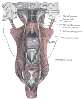
 Dissection of the muscles of the palate from behind. | |
| Details | |
| Identifiers | |
| Latin | aponeurosis palatina |
| Anatomical terminology | |
The palatine aponeurosis a thin, firm, fibrous lamella[1] which gives strength[2] and support to soft palate.[3] It serves as the insertion for the tensor veli palatini and levator veli palatini, and the origin for the musculus uvulae, palatopharyngeus, and palatoglossus.[4]
The palatine aponeurosis is attached to the posterior margin of the hard palate.[2][5] It is thicker anteriorly and thiner posteriorly. Posteriorly, it blends with the posterior muscular part of the soft palate. Posteroinferiorly, it presents a cruved free margin from which the uvula is suspended.[2] Laterally, it is continuous with the pharyngeal aponeurosis.[1]
See also
References
- ↑ 1.0 1.1 Gray, Henry (1918). Gray's Anatomy (20th ed.). pp. 1139. https://archive.org/details/anatomyofhumanbo1918gray/page/659/mode/2up?view=theater.
- ↑ 2.0 2.1 2.2 Moore, Keith L.; Dalley, Arthur F.; Agur, Anne M. R. (2017). Essential Clinical Anatomy. Lippincott Williams & Wilkins. pp. 943. ISBN 978-1496347213.
- ↑ Sauerland, Eberhardt K.; Patrick W. Tank; Tank, Patrick W. (2005). Grant's dissector. Hagerstown, MD: Lippincott Williams & Wilkins. p. 199. ISBN 0-7817-5484-4. https://archive.org/details/grantsdissector00tank_714.
- ↑ Anne M. R. Agur; Moore, Keith L. (2006). Essential Clinical Anatomy (Point (Lippincott Williams & Wilkins)). Hagerstown, MD: Lippincott Williams & Wilkins. p. 553. ISBN 0-7817-6274-X.
- ↑ Gray, Henry (1918). Gray's Anatomy (20th ed.). pp. 1139. https://archive.org/details/anatomyofhumanbo1918gray/page/1139/mode/2up?view=theater.
 |

