Biology:Mass cytometry

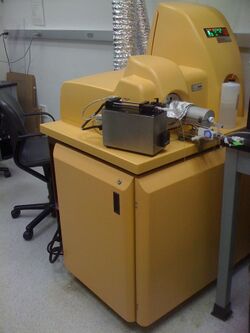
Mass cytometry is a mass spectrometry technique based on inductively coupled plasma mass spectrometry and time of flight mass spectrometry used for the determination of the properties of cells (cytometry).[1][2] In this approach, antibodies are conjugated with isotopically pure elements, and these antibodies are used to label cellular proteins. Cells are nebulized and sent through an argon plasma, which ionizes the metal-conjugated antibodies. The metal signals are then analyzed by a time-of-flight mass spectrometer. The approach overcomes limitations of spectral overlap in flow cytometry by utilizing discrete isotopes as a reporter system instead of traditional fluorophores which have broad emission spectra.[3]
Commercialization
Tagging technology and instrument development occurred at the University of Toronto and DVS Sciences, Inc.[1][4] CyTOF (cytometry by time of flight) was initially commercialized by DVS Sciences in 2009. In 2014, Fluidigm acquired DVS Sciences [5] to become a reference company in single cell technology.[6] In 2022 Fluidigm received a capitol infusion and changed its name to Standard BioTools. [7] The CyTOF, CyTOF2, Helios (CyTOF3) and CyTOF XT[8](4th generation) have been commercialized up to now. Fluidigm sells a variety of commonly used metal-antibody conjugates, and an antibody conjugation kit.
Imaging Mass Cytometry (IMC)
Imaging mass cytometry (IMC) is a relatively new imaging technique, emerged from previously available CyTOF technology (cytometry by time of flight), that combines mass spectrometry with UV laser ablation to generate pseudo images of tissue samples.[9][10] This approach adds spatial resolution to the data, which enables simultaneous analysis of multiple cell markers at subcellular resolution and their spatial distribution in tissue sections.[9][11] The IMC approach, in the same way as CyTOF, relies on detection of metal-tagged antibodies using time-of-flight mass spectrometry, allowing for quantification of up to 40 markers simultaneously.[12][13]
Data analysis
CyTOF mass cytometry data is recorded in tables that list, for each cell, the signal detected per channel, which is proportional to the number of antibodies tagged with the corresponding channel's isotope bound to that cell. These data are formatted as FCS files, which are compatible with traditional flow cytometry software. Due to the high-dimensional nature of mass cytometry data, novel data analysis tools have been developed as well.[14]
Imaging Mass Cytometry data analysis has its specificity due to different nature of data obtained. In terms of data analysis, both IMC and CyTOF generate large datasets with high dimensionality that require specialized computational methods for analysis. However, data generated by IMC can be more challenging to analyze due to additional data complexity and need for specific tools and pipelines specific for digital image analysis, whereas the data generated by CyTOF is generally analyzed using conventional flow cytometry software. A comprehensive overview of IMC data analysis techniques has been given by Milosevic in.[15]
Advantages and disadvantages
Advantages include minimal overlap in metal signals meaning the instrument is theoretically capable of detecting 100 parameters per cell, entire cell signaling networks can be inferred organically without reliance on prior knowledge, and one well-constructed experiment produces large amounts of data.[3]
Disadvantages, in the case of CyTOF, include the practical flow rate is around 500 cells per second versus several thousand in flow cytometry and current reagents available limit cytometer use to around 50 parameters per cell. Additionally, mass cytometry is a destructive method and cells cannot be sorted for further analysis. In the case of IMC, the resolution of the data is relatively low (1μm2/pixel), the technique is as well destructive, acquiring of the data is also very slow, and it requires specialized expensive equipment and expertise.
Applications
Mass cytometry has research applications in medical fields including immunology, hematology, and oncology. It has been used in studies of hematopoiesis,[16] cell cycle,[17] cytokine expression, and differential signaling responses.
MC has been used in various research fields, such as cancer biology, immunology, and neuroscience, to provide a more comprehensive understanding of tissue architecture and cellular interactions.[18][19][20][21][22][23]
References
- ↑ 1.0 1.1 Dmitry Bandura; Vladimir Baranov; Olga Ornatsky; Scott D. Tanner; Kinach R; Lou X; Pavlov S; Vorobiev S et al. (2009). "Mass Cytometry: Technique for Real Time Single Cell Multitarget Immunoassay Based on Inductively Coupled Plasma Time-of-Flight Mass Spectrometry". Analytical Chemistry 81 (16): 6813–6822. doi:10.1021/ac901049w. PMID 19601617.
- ↑ Di Palma, Serena; Bodenmiller, Bernd (2015). "Unraveling cell populations in tumors by single-cell mass cytometry". Current Opinion in Biotechnology 31: 122–129. doi:10.1016/j.copbio.2014.07.004. ISSN 0958-1669. PMID 25123841.
- ↑ 3.0 3.1 Spitzer, Matthew H.; Nolan, Garry P. (2016). "Mass Cytometry: Single Cells, Many Features". Cell 165 (4): 780–791. doi:10.1016/j.cell.2016.04.019. ISSN 0092-8674. PMID 27153492.
- ↑ Ornatsky, O; Bandura D; Baranov V; Nitz M; Winnick MA; Tanner S (30 September 2010). "Highly multiparametric analysis by mass cytometry". Journal of Immunological Methods 361 (1–2): 1–20. doi:10.1016/j.jim.2010.07.002. PMID 20655312.
- ↑ "Fluidigm | Press Releases | FLUIDIGM TO ACQUIRE DVS SCIENCES". https://www.fluidigm.com/press/fluidigm-to-acquire-dvs-sciences. Retrieved 2015-11-11.
- ↑ "An Open Letter to Customers of Fluidigm and DVS Sciences, Inc.". Fluidigm and DVS Sciences, Inc.. 13 February 2014. http://www.dvssciences.com/uploads/Open_Letter_to_Customers_Feb_13_2014.pdf. Retrieved 4 July 2014.
- ↑ "Standard BioTools| Press Releases | Fluigigm Capital Infusion". https://www.globenewswire.com/news-release/2022/01/24/2371694/0/en/Fluidigm-Announces-250-Million-Strategic-Capital-Infusion-from-Casdin-Capital-and-Viking-Global-Investors-and-Rebranding-to-Standard-BioTools-Inc.html. Retrieved 2022-01-22.
- ↑ "CyTOF XT". 2021-05-21. https://investors.fluidigm.com/news-releases/news-release-details/next-generation-cytof-xt-redefines-cytometry-advances-automation.
- ↑ 9.0 9.1 Giesen, Charlotte; Wang, Hao A O; Schapiro, Denis; Zivanovic, Nevena; Jacobs, Andrea; Hattendorf, Bodo; Schüffler, Peter J; Grolimund, Daniel et al. (2014). "Highly multiplexed imaging of tumor tissues with subcellular resolution by mass cytometry" (in en). Nature Methods 11 (4): 417–422. doi:10.1038/nmeth.2869. ISSN 1548-7091. PMID 24584193. http://www.nature.com/articles/nmeth.2869.
- ↑ Chang, Qing; Ornatsky, Olga I.; Siddiqui, Iram; Loboda, Alexander; Baranov, Vladimir I.; Hedley, David W. (2017). "Imaging Mass Cytometry" (in en). Cytometry Part A 91 (2): 160–169. doi:10.1002/cyto.a.23053. PMID 28160444.
- ↑ Schapiro, Denis; Jackson, Hartland W; Raghuraman, Swetha; Fischer, Jana R; Zanotelli, Vito R T; Schulz, Daniel; Giesen, Charlotte; Catena, Raúl et al. (2017). "histoCAT: analysis of cell phenotypes and interactions in multiplex image cytometry data" (in en). Nature Methods 14 (9): 873–876. doi:10.1038/nmeth.4391. ISSN 1548-7091. PMID 28783155.
- ↑ Ijsselsteijn, Marieke E.; van der Breggen, Ruud; Farina Sarasqueta, Arantza; Koning, Frits; de Miranda, Noel F. C. C. (2019-10-29). "A 40-Marker Panel for High Dimensional Characterization of Cancer Immune Microenvironments by Imaging Mass Cytometry". Frontiers in Immunology 10: 2534. doi:10.3389/fimmu.2019.02534. ISSN 1664-3224. PMID 31736961.
- ↑ Han, Guojun; Chen, Shih-Yu; Gonzalez, Veronica D.; Zunder, Eli R.; Fantl, Wendy J.; Nolan, Garry P. (2017). "Atomic mass tag of bismuth-209 for increasing the immunoassay multiplexing capacity of mass cytometry" (in en). Cytometry Part A 91 (12): 1150–1163. doi:10.1002/cyto.a.23283. ISSN 1552-4922. PMID 29205767.
- ↑ Krishnaswamy, Smita; Spitzer, Matthew; Mingueneau, Michael; Bendall, Sean; Litvin, Oren; Stone, Erica; Pe'er, Dana; Nolan, Garry (28 Nov 2014). "Conditional density-based analysis of T cell signaling in single-cell data". Science 346 (6213): 1250689. doi:10.1126/science.1250689. PMID 25342659.
- ↑ Milosevic, Vladan (2023). "Different approaches to Imaging Mass Cytometry data analysis" (in en). Bioinformatics Advances 3: vbad046. doi:10.1093/bioadv/vbad046. ISSN 2635-0041. PMID 37092034. PMC 10115470. https://academic.oup.com/bioinformaticsadvances/advance-article/doi/10.1093/bioadv/vbad046/7100350.
- ↑ "Single-cell mass cytometry of differential immune and drug responses across a human hematopoietic continuum". Science 332 (6030): 687–96. 2011. doi:10.1126/science.1198704. PMID 21551058. Bibcode: 2011Sci...332..687B.
- ↑ Behbehani, Gregory K.; Bendall, Sean C.; Clutter, Matthew R.; Fantl, Wendy J.; Nolan, Garry P. (2012-07-01). "Single-cell mass cytometry adapted to measurements of the cell cycle". Cytometry Part A 81 (7): 552–566. doi:10.1002/cyto.a.22075. ISSN 1552-4930. PMID 22693166.
- ↑ Damond, Nicolas; Engler, Stefanie; Zanotelli, Vito R.T.; Schapiro, Denis; Wasserfall, Clive H.; Kusmartseva, Irina; Nick, Harry S.; Thorel, Fabrizio et al. (2019). "A Map of Human Type 1 Diabetes Progression by Imaging Mass Cytometry" (in en). Cell Metabolism 29 (3): 755–768.e5. doi:10.1016/j.cmet.2018.11.014. PMID 30713109.
- ↑ Ramaglia, Valeria; Sheikh-Mohamed, Salma; Legg, Karen; Park, Calvin; Rojas, Olga L; Zandee, Stephanie; Fu, Fred; Ornatsky, Olga et al. (2019-08-01). "Multiplexed imaging of immune cells in staged multiple sclerosis lesions by mass cytometry" (in en). eLife 8: e48051. doi:10.7554/eLife.48051. ISSN 2050-084X. PMID 31368890.
- ↑ Jackson, Hartland W.; Fischer, Jana R.; Zanotelli, Vito R. T.; Ali, H. Raza; Mechera, Robert; Soysal, Savas D.; Moch, Holger; Muenst, Simone et al. (2020-02-27). "The single-cell pathology landscape of breast cancer" (in en). Nature 578 (7796): 615–620. doi:10.1038/s41586-019-1876-x. ISSN 0028-0836. PMID 31959985. Bibcode: 2020Natur.578..615J. http://www.nature.com/articles/s41586-019-1876-x.
- ↑ Moldoveanu, Dan; Ramsay, LeeAnn; Lajoie, Mathieu; Anderson-Trocme, Luke; Lingrand, Marine; Berry, Diana; Perus, Lucas J.M.; Wei, Yuhong et al. (2022). "Spatially mapping the immune landscape of melanoma using imaging mass cytometry" (in en). Science Immunology 7 (70): eabi5072. doi:10.1126/sciimmunol.abi5072. ISSN 2470-9468. PMID 35363543. https://www.science.org/doi/10.1126/sciimmunol.abi5072.
- ↑ Hoch, Tobias; Schulz, Daniel; Eling, Nils; Gómez, Julia Martínez; Levesque, Mitchell P.; Bodenmiller, Bernd (2022). "Multiplexed imaging mass cytometry of the chemokine milieus in melanoma characterizes features of the response to immunotherapy" (in en). Science Immunology 7 (70): eabk1692. doi:10.1126/sciimmunol.abk1692. ISSN 2470-9468. PMID 35363540. https://www.science.org/doi/10.1126/sciimmunol.abk1692.
- ↑ Liu, Zehan; Xun, Jing; Liu, Shuangqing; Wang, Botao; Zhang, Aimin; Zhang, Lanqiu; Wang, Ximo; Zhang, Qi (2022). "Imaging mass cytometry: High-dimensional and single-cell perspectives on the microenvironment of solid tumours" (in en). Progress in Biophysics and Molecular Biology 175: 140–146. doi:10.1016/j.pbiomolbio.2022.10.003. PMID 36252872. https://linkinghub.elsevier.com/retrieve/pii/S0079610722001031.
 |
