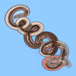
Biology:Nippostrongylus brasiliensis
| Nippostrongylus brasiliensis | |
|---|---|

| |
| Scientific classification | |
| Domain: | Eukaryota |
| Kingdom: | Animalia |
| Phylum: | Nematoda |
| Class: | Chromadorea |
| Order: | Rhabditida |
| Suborder: | Strongylida |
| Superfamily: | Trichostrongyloidea |
| Family: | Heligmonellidae |
| Subfamily: | Nippostrongylinae |
| Genus: | Nippostrongylus |
| Species: | N. brasiliensis
|
| Binomial name | |
| Nippostrongylus brasiliensis (Travassos, 1914)
| |
Nippostrongylus brasiliensis is a gastrointestinal roundworm that infects rodents, primarily rats. This worm is a widely studied parasite due to its simple lifecycle and its ability to be used in animal models. Its lifecycle similar to the human hookworms Necator americanus and Ancylostoma duodenale which includes five molting stages to become sexually mature.
Lifecycle
Eggs located within the soil release motile, free-living worms that must moult twice (L1 and L2) to develop into their infective L3 stage. This L3 stage can penetrate through intact skin in as little as 4 hours. Once inside the host, the worms invade the venous circulation and are carried into the lungs, where they become trapped in the capillaries. When the worms mature into the L4 stage, they rupture the capillaries and are released into the alveoli, where they are coughed up and swallowed. They then reach the small intestines 3–4 days after the initial infection. The worms become adults after the final molt into the L5 stage, where they begin laying eggs on day 6 of infection.[1] The eggs are passed out of the host through feces and the cycle starts all over again.
N. brasiliensis is adapted to infecting rats, so can continue laying eggs for prolonged periods of time. The immune response of mice, however, leads to cessation of egglaying by day 8 and adults are expelled by day 10.[2]
Animal model
N. brasiliensis provides a valuable lab model in determining the migration pathway through the host. The lifecycle of N. brasiliensis can be passed through lab mice. The availability of inbred and mutant mouse strains can be advantageous when examining the genetic basis of murine susceptibility and resistance to infection.[3] Animal models of N. brasiliensis infections can lead to a better understanding of the basic biology of the immune response and protective immunity. For instance, they can provide the model for induction and maintenance of Th2 type immune responses and exhibit all the characteristics for eosinophilia, mastocytosis, mucus production, and CD4 T cell-dependent IgE production.[4] An infection model of N. brasiliensis has been used to determine that at least two distinct Th2-type immune responses occur - one that is TSLP-dependent, and one that is type-1 interferon-dependent.[5]
Symptoms and diseases
Lab mice previously infected with N. brasiliensis develop massive emphysema with dilation of distal airspaces due to the loss of alveolar septa; N. brasiliensis infection can result in deterioration of the lung, destruction to the alveoli, and long-term airway hyperresponsiveness, which is consistent with emphysema and chronic obstructive pulmonary disorder (COPD). This infection can lead to a chronic low level hemorrhaging of the lung. The damage to the lung tissue can result in the development of COPD and emphysema.[6] Among respiratory problems, a N. brasiliensis infection can also result in the loss of both body mass and red blood cell density. Biphasic anorexia is also prevalent in laboratory rats infected with this parasite. The first phase coincides with the parasites invasion of the lung and the second phase occurs when the parasite matures inside the intestine.[7]
Treatment
Tetramisole loaded into zeolite is more effective at killing adults of N. brasiliensis in rats than tetramisole alone.[8]
See also
References
- ↑ Locksley, Richard M., and Miranda Robertson. "11.4." Immunity: The Immune Response in Infectious and Inflammatory Disease. By Anthony L. DeFranco. N.p.: New Science, 2007. 274-75. Print.
- ↑ Locksley, Richard M., and Miranda Robertson. "11.4." Immunity: The Immune Response in Infectious and Inflammatory Disease. By Anthony L. DeFranco. N.p.: New Science, 2007. 274-75. Print.
- ↑ Stadnyk, A.W., P.J. McElroy, J. Gauldie, and A.D. Befus. 1990. Characterization of Nippostrongylus brasiliensis infection in different strains of mice. The Journal of Parasitology 76:377-382.
- ↑ Camberis, M., G. Le Gros, and J. Urban Jr. 2003. Animal model of Nippostrongylus brasiliensis and Heligmosmoides polygyrus. Current Protocols in Immunology Chapter 19.
- ↑ Lisa M. Connor, Shiau-Choot Tang, Emmanuelle Cognard, Sotaro Ochiai, Kerry L. Hilligan, Samuel I. Old, Christophe Pellefigues, Ruby F. White, Deepa Patel, Adam Alexander T. Smith, David A. Eccles, Olivier Lamiable, Melanie J. McConnell, Franca Ronchese. 2017. Th2 responses are primed by skin dendritic cells with distinct transcriptional profiles. Journal of Experimental Medicine 214 (1): 125-142; DOI: 10.1084/jem.20160470
- ↑ Marsland, B.J., M. Kurrer, R. Reissmann, N.L. Harris, and M. Kopf. 2008. Nippostrongylus brasiliensis infection leads to the development of emphysema associated with the induction of alternatively activated macrophages. European Journal of Immunology 38:479-488.
- ↑ Mercer, J.G., P.I. Mitchell, K.M. Moar, A. Bissett, S. Geissler, K. Bruce, and L.H. Chappell. 2000. Anorexia in rats infected with the nematode, Nippostrongylus brasiliensis: experimental manipulations. Parasitology 120:641-647.
- ↑ Shaker, S.K., A. Dyer, and D.M. Storey. 1992. Treatment of Nippostrongylus brasiliensis in normal and SPF rats using tetramisole loaded with zeolite. Journal of Helminthology 66:288-292.
Wikidata ☰ Q5267184 entry
 |

