Biology:Splenogonadal fusion
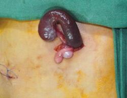
Splenogonadal fusion is a rare congenital malformation that results from an abnormal connection between the primitive spleen and gonad during gestation. A portion of the splenic tissue then descends with the gonad. Splenogonadal fusion has been classified into two types: continuous, where there remains a connection between the main spleen and gonad; and discontinuous, where ectopic splenic tissue is attached to the gonad, but there is no connection to the orthotopic spleen. Patients with continuous splenogonadal fusion frequently have additional congenital abnormalities, most commonly cryptorchidism.[1]
The anomaly was first described in 1883 by Bostroem.[2] Since then more than 150 cases of splenogonadal fusion have been documented.[3] The condition is considered benign.[4] A few cases of testicular neoplasm have been reported in association with splenogonadal fusion.[5][6] The reported cases have occurred in patients with a history of cryptorchidism, which is associated with an elevated risk of neoplasm.[6]
Splenogonadal fusion occurs with a male-to-female ratio of 16:1, and is seen nearly exclusively on the left side.[3] The condition remains a diagnostic challenge, but preoperative consideration of the diagnosis may help avoid unnecessary orchiectomy. On scrotal ultrasound, ectopic splenic tissue may appear as an encapsulated homogeneous extratesticular mass, isoechoic with the normal testis. Subtle hypoechoic nodules may be present in the mass.[7] The presence of splenic tissue may be confirmed with a technetium-99m sulfur colloid scan.[8]
References
- ↑ Kocher, NJ; Tomaszewski, JJ; Parsons, RB; Cronson, BR; Altman, H; Kutikov, A (Jan 2014). "Splenogonadal fusion: a rare etiology of solid testicular mass". Urology 83 (1): e1-2. doi:10.1016/j.urology.2013.09.019. PMID 24200197.
- ↑ Khairat, AB; Ismail, AM (Aug 2005). "Splenogonadal fusion: case presentation and literature review". Journal of Pediatric Surgery 40 (8): 1357–60. doi:10.1016/j.jpedsurg.2005.05.027. PMID 16080949.
- ↑ 3.0 3.1 Varma, DR; Sirineni, GR; Rao, MV; Pottala, KM; Mallipudi, BV (Sep 2007). "Sonographic and CT features of splenogonadal fusion". Pediatric radiology 37 (9): 916–9. doi:10.1007/s00247-007-0526-x. PMID 17581747.
- ↑ Lin, CS; Lazarowicz, JL; Allan, RW; Maclennan, GT (Jul 2010). "Splenogonadal fusion". The Journal of Urology 184 (1): 332–3. doi:10.1016/j.juro.2010.04.013. PMID 20488470.
- ↑ Imperial, SL; Sidhu, JS (Oct 2002). "Nonseminomatous germ cell tumor arising in splenogonadal fusion". Archives of Pathology & Laboratory Medicine 126 (10): 1222–5. doi:10.1043/0003-9985(2002)126<1222:NGCTAI>2.0.CO;2. PMID 12296764.
- ↑ 6.0 6.1 "Splenogonadal fusion and testicular cancer: case report and review of the literature". Einstein (Sao Paulo, Brazil) 10 (1): 92–5. Jan–Mar 2012. doi:10.1590/s1679-45082012000100019. PMID 23045834.
- ↑ Ferrón, SA; Arce, JD (Dec 2013). "Discontinuous splenogonadal fusion: new sonographic findings". Pediatric radiology 43 (12): 1652–5. doi:10.1007/s00247-013-2730-1. PMID 23754542.
- ↑ Guarin, U; Dimitrieva, Z; Ashley, SJ (Oct 1975). "Splenogonadal fusion-a rare congenital anomaly demonstrated by 99Tc-sulfur colloid imaging: case report". Journal of Nuclear Medicine 16 (10): 922–4. PMID 240914.
 |


