Engineering:Brewster angle microscope


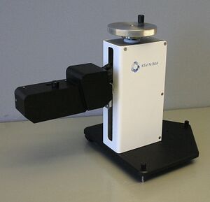
A Brewster angle microscope (BAM) is a microscope for studying thin films on liquid surfaces, most typically Langmuir films.[1] In a Brewster angle microscope, both the microscope and a polarized light source are aimed towards a liquid surface at that liquid's Brewster angle, in such a way for the microscope to catch an image of any light reflected from the light source via the liquid surface. Because there is no p-polarized reflection from the pure liquid when both are angled towards it at the Brewster angle, light is only reflected when some other phenomenon such as a surface film affects the liquid surface.[2] The technique was first introduced in 1991.[3]
Applications
Brewster angle microscopes enable the visualization of Langmuir monolayers or adsorbate films at the air-water interface for example as a function of packing density.[1] They can be used either to study the properties of the Langmuir layer, or to indicate a suitable deposition pressure for Langmuir-Blodgett (LB) deposition. They can be used for example in the LB deposition of nanoparticles. Applications include:[4]
Monolayer/film homogeneity. When combined with a Langmuir-Blodgett Trough, observation can be performed during compression/expansion at known surface pressures.
Optimizing the deposition parameters. Selecting optimal deposition pressure and other deposition parameters for LB coating.
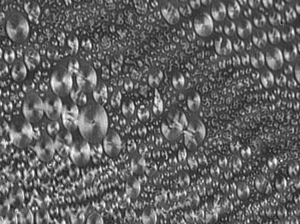
Monolayer/film behavior. Observing phase changes, phase separation, domain size, shape and packing.
Monitoring of surface reactions. Photochemical reactions, polymerizations and enzyme kinetics can be followed in real time.
Monitoring and detection of surface active materials. For example, protein adsorption and nanoparticle flotation.
Lee et al.[5] used a Brewster angle microscope to study optimal deposition parameters for Fe3O4 nanoparticles.
Daear et al.[6] have written a recent review on the usage of BAMs in biological applications.
See also
References
- ↑ 1.0 1.1 Daear, Weiam; Mahadeo, Mark; Prenner, Elmar J. (2017-10-01). "Applications of Brewster angle microscopy from biological materials to biological systems". Biochimica et Biophysica Acta (BBA) - Biomembranes 1859 (10): 1749–1766. doi:10.1016/j.bbamem.2017.06.016. ISSN 0005-2736. PMID 28655618.
- ↑ "Brewster Angle Microscopy - Biolin Scientific" (in en-US). Biolin Scientific. http://www.biolinscientific.com/technology/brewster-angle-microscopy/.
- ↑ M. A. Cohen Stuart; R. A. J. Wegh. "Design and Testing of a Low-Cost and Compact Brewster Angle Microscope". http://www.tu-chemnitz.de/physik/OSMP/Soft/ws0607_ue05a.pdf. Langmuir, Vol. 12, No. 11, 1996. p. 2863
- ↑ "Imaging the Structure of Thin Films: Brewster Angle Microscopy". http://www.biolinscientific.com/zafepress.php?url=%2Fpdf%2FKSV%20NIMA%2FApplication%20Notes%2FKN-AN-09-Brewster-angle-microscopy-thin-films.pdf.
- ↑ Lee, Don Keun; Kim, Young Hwan; Kim, Chang Woo; Cha, Hyun Gil; Kang, Young Soo (2007-08-01). "Vast Magnetic Monolayer Film with Surfactant-Stabilized Fe3O4 Nanoparticles Using Langmuir−Blodgett Technique". The Journal of Physical Chemistry B 111 (31): 9288–9293. doi:10.1021/jp072612c. ISSN 1520-6106. PMID 17636981.
- ↑ Daear, Weiam; Mahadeo, Mark; Prenner, Elmar J. (2017). "Applications of Brewster angle microscopy from biological materials to biological systems". Biochimica et Biophysica Acta (BBA) - Biomembranes 1859 (10): 1749–1766. doi:10.1016/j.bbamem.2017.06.016. PMID 28655618.
External links
 |
