Medicine:Pharyngeal plexus of vagus nerve
| Pharyngeal plexus of vagus nerves | |
|---|---|
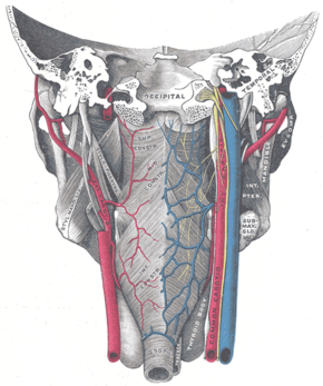
 Muscles of the pharynx, viewed from behind, together with the associated vessels and nerves. (Pharyngeal plexus visible but not labeled.) | |
| Details | |
| Identifiers | |
| Latin | plexus pharyngeus nervi vagi |
| Anatomical terms of neuroanatomy | |
The pharyngeal plexus is a nerve plexus located upon the outer surface of the pharynx. It contains a motor component (derived from the vagus nerve (cranial nerve X)), a sensory component (derived from the glossopharyngeal nerve (cranial nerve IX)), and sympathetic component (derived from the superior cervical ganglion).[1]
The plexus provides motor innervation to most muscles of the soft palate (all but the tensor veli palatini muscle) and most muscles of the pharynx (all but the stylopharyngeus muscle).[1] The larynx meanwhile receives motor innervation from the vagus nerve (CN X) via its external branch of the superior laryngeal nerve and its recurrent laryngeal nerve, and not through the pharyngeal plexus.[citation needed]
Anatomy
The pharyngeal plexus occurs upon the outer surface of the pharynx - especially superficial to the middle pharyngeal constrictor muscle.[1]
Afferents
It has the following components:[2]
- Motor – pharyngeal branch of vagus nerve (CN X) which arises from the superior portion of the inferior ganglion of vagus nerve, with the neuron cell bodies of its axons residing in the nucleus ambiguus. The pharyngeal branch ramifies upon reaching the superior border of the middle pharyngeal constrictor muscle. It is unclear whether the cranial root of accessory nerve (CN XI)[1]
- Sensory – pharyngeal branches of glossopharyngeal nerve.
- Sympathetic vasomotor – efferent fibres of the superior cervical ganglion.
Because the cranial part of accessory nerve (CN XI) leaves the jugular foramen as a part of the CN X, it is sometimes considered part of the plexus as well.[2]
Efferents/distribution
En route to their target tissues, outgoing fibres from the pharyngeal plexus at first either ascend upon the superior pharyngeal constrictor muscle or descend upon the inferior pharyngeal constrictor muscle, then ramify within the pharyngeal muscular layer and mucous membrane.[1]
Motor
The pharyngeal plexus, with fibers from CN IX, CN X, and cranial part of CN XI, innervates all the muscles of the pharynx (except stylopharyngeus, which is innervated directly by a branch of CN IX).[dubious ][citation needed]
This includes the following muscles: palatopharyngeus, palatoglossus, musculus uvulae, the pharyngeal constrictors, salpingopharyngeus plus others.[citation needed]
Sensory
The pharyngeal plexus provides sensory innervation to most of the pharynx;[1] it provides sensory innervation to the oropharynx and laryngopharynx from CN IX and CN X. (The nasopharynx above the pharyngotympanic tube and the torus tubarius is innervated by CN V2).[citation needed]
Sympathetic
See also
- Superior cervical ganglion
Additional images
References
- ↑ Jump up to: 1.0 1.1 1.2 1.3 1.4 1.5 Standring, Susan (2020). Gray's Anatomy: The Anatomical Basis of Clinical Practice (42th ed.). New York. pp. 713. ISBN 978-0-7020-7707-4. OCLC 1201341621. https://www.worldcat.org/oclc/1201341621.
- ↑ Jump up to: 2.0 2.1 Jerry L. Gadd; James L. Oschman; Hiatt, James L.; Leslie P. Gartner; Gartner, Leslie P. (2001). Textbook of head and neck anatomy. Hagerstown, MD: Lippincott Williams & Wilkins. p. 229. ISBN 0-7817-2166-0.
 |


