Medicine:Temporal styloid process
| Temporal styloid process | |
|---|---|
 Right side of the skull. Styloid process shown in red | |
 Right temporal bone and mandible (styloid process labeled at bottom) | |
| Details | |
| Identifiers | |
| Latin | processus styloideus ossis temporalis |
| Anatomical terms of bone | |
The temporal styloid process is a slender bony process of the temporal bone extending downward and forward from the undersurface of the temporal bone[1] just below the ear. The styloid process gives attachments to several muscles, and ligaments.
Structure
The styloid process is a slender and pointed bony process of the temporal bone projecting anteroinferiorly from the inferior surface of the temporal bone[1] just below the ear. Its length normally ranges from just under 3 cm to just over 4 cm. It is usually nearly straight, but may be curved in some individuals.[1]
Its proximal (tympanohyal) part is ensheathed by the tympanic part of the temporal bone (vaginal process), whereas its distal (stylohyal) part gives attachment to several structures.[1]
Attachments
The styloid process gives attachments to several muscles, and ligaments.[1] It serves as an anchor point for several muscles associated with the tongue and larynx. * stylohyoid ligament[2]
Relations
The parotid gland is situated laterally to the styloid process, the external carotid artery passes by its apex, the facial nerve crosses its base, and the attachment of the stylopharyngeus muscle separates it from the internal jugular vein medially.[1]
Development
The styloid process arises from endochondral ossification of the cartilage from the second pharyngeal arch (Reichert's cartilage).[3] It arises from fusion of dorsal and ventral components of the arch.[2]
Clinical significance
In other animals
The styloid process is present across mammals.[2]
The origin of the structure that would become the styloid process is in Reichert's cartilage, which makes up the second branchial arch in ancestral vertebrates. In gnathostomes, this cartilage separated into two discrete elements: the hyomandibula and the ceratohyal.[2] The hyomandibula in turn evolved into two discrete elements in basal amniotes: the columella and the extracolumella (believed to be equivalent to the mammalian tympanohyal),[4] while the ceratohyal evolved into the mammalian stylohyal. Developmental evidence suggests that the styloid process evolved from a fusion of the tympanohyal and the stylohyal.[2]
Additional images
-
 Animation. Temporal styloid process shown in red.
-
 Left temporal bone.
-
 Inferior surface of left temporal bone. Styloid process shown in red.
-
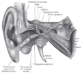 External and middle ear, opened from the front. Right side. (Label for styloid process is bottom center.)
-
 Left temporal bone. Outer surface. (Styloid process visible at center bottom.)
-
 Articulation of the mandible. Medial aspect.
-
 Extrinsic muscles of the tongue. Left side.
-
 Dissection of the muscles of the palate from behind.
-
 Styloid process. Base of skull.


References
- ↑ 1.0 1.1 1.2 1.3 1.4 1.5 Gray's anatomy : the anatomical basis of clinical practice. Susan Standring (Forty-second ed.). [New York]. 2021. pp. 737. ISBN 978-0-7020-7707-4. OCLC 1201341621.
- ↑ 2.0 2.1 2.2 2.3 2.4 Kitazawa, Taro; Fujisawa, Kou; Narboux-Nême, Nicolas; Arima, Yuichiro; Kawamura, Yumiko; Inoue, Tsuyoshi; Wada, Youichiro; Kohro, Takahide et al. (2015-06-15). "Distinct effects of Hoxa2 overexpression in cranial neural crest populations reveal that the mammalian hyomandibular-ceratohyal boundary maps within the styloid process". Developmental Biology 402 (2): 162–174. doi:10.1016/j.ydbio.2015.04.007. ISSN 0012-1606. https://www.sciencedirect.com/science/article/pii/S0012160615001876.
- ↑ Rodríguez-Vázquez, J. F.; Mérida-Velasco, J. R.; Verdugo-López, S.; Sánchez-Montesinos, I.; Mérida-Velasco, J. A. (2006). "Morphogenesis of the second pharyngeal arch cartilage (Reichert's cartilage) in human embryos" (in en). Journal of Anatomy 208 (2): 179–189. doi:10.1111/j.1469-7580.2006.00524.x. ISSN 1469-7580. PMID 16441562. PMC 2100189. https://onlinelibrary.wiley.com/doi/abs/10.1111/j.1469-7580.2006.00524.x.
- ↑ Allin, Edgar F.; Hopson, James A. (1992), "Evolution of the Auditory System in Synapsida (“Mammal-Like Reptiles” and Primitive Mammals) as Seen in the Fossil Record", in Webster, Douglas B.; Popper, Arthur N.; Fay, Richard R. (in en), The Evolutionary Biology of Hearing, New York, NY: Springer, pp. 587–614, doi:10.1007/978-1-4612-2784-7_37, ISBN 978-1-4612-2784-7, https://doi.org/10.1007/978-1-4612-2784-7_37, retrieved 2026-07-20
This article incorporates text in the public domain from the 20th edition of Gray's Anatomy (1918)
External links
- Anatomy photo:22:os-0407 at the SUNY Downstate Medical Center
- "Anatomy diagram: 25420.000-1". Roche Lexicon - illustrated navigator. Elsevier. http://www.tk.de/rochelexikon/pics/s25420.000-1.html.
- "Anatomy diagram: 34257.000-1". Roche Lexicon - illustrated navigator. Elsevier. http://www.tk.de/rochelexikon/pics/s34257.000-1.html.
 |  |


