Multiple scattering low coherence interferometry
Multiple scattering low coherence interferometry (ms/LCI) is an imaging technique that relies on analyzing multiply scattered light in order to capture depth-resolved images from optical scattering media. With current applications primarily in medical imaging, has the advantage of a higher range since forward scattered light attenuates less with depth when compared to the specularly reflected light that is assessed in more conventional imaging methods such as optical coherence tomography.[1] This allows ms/LCI to image through up to 90 mean free scattering paths, compared to roughly 27 scattering MFPs in OCT and 1–2 scattering MFPs in confocal microscopy.[2]
Design
Time-domain implementation
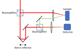
Early implementations of ms/LCI were in the time domain using lock-in detection in order to take advantage of long scanning depths as well narrow detection bandwidths.[2] As in traditional OCT, the beam interference coherence gates the light in order to filter out photons that have not traveled a sufficient path length. The use of distinct, non-intersecting illumination and collection beams allows for time insensitive triangulation of the light that also considers physical depth penetration into the media in order to reject diffuse backscattering light such that only forward scattered photons are analyzed. Unlike traditional OCT, the measured forward scattered light is diverged by a lens in order to isolate the angular component to be compared with the reference arm. Due to the dominance of forward scattering photons at deeper penetration depths, this technique enjoys superior imaging depth and high detection throughput but suffers from long signal acquisition time and poor spatial resolution inherent to time-domain techniques.
Spectral-domain implementation
By adapting techniques used in spectroscopic OCT, ms/LCI can be done in the spectral domain in order to provide faster acquisition time and various multispectral capabilities, including the use of localized contrast agents.[1] As in OCT, enhanced depth imaging is used to place the zero-path delay point behind the focal volume in order to increase sensitivity. A broadband supercontinuum laser source as well as a customized spectrometer detector are implemented in order to access the spectral domain, and a short-time Fourier transform method is used to process the interferograms as in spectroscopic OCT.

Applications
Because of enhanced depth penetration, this technique's most promising applications lie in resolving features that exist deeper in tissue than other techniques such as OCT are capable of doing. Studies have determined ms/LCI's feasibility in a variety of clinical applications, including assessing burn injuries, as well as in vivo imaging of rat skin with superior depth results when compared to OCT.[3]
Considerations
Spatial resolution
Photons originating from the focal plane detected by ms/LCI will undergo multiple scattering events which causes extra travel time and resulting longer path lengths than the signal would otherwise indicate. This axial shift will make photons appear like they appear deeper from the sample than they actually are.
The presence of multiple scattering events causes a distribution of path lengths that intrinsically blurs the image, resulting in a maximum millimeter-scale resolution which is substantially poorer than OCT which operates at a micrometer-scale resolution.
Because of anisotropic propagation of light in tissue, the lateral profile will spread out slowly relative to axial profile, resulting in an elongated image.
Spectral-domain ms/LCI uniquely needs to account for the wavelength dependence of scattering when interpreting the reflectance depth profile as well as the transfer of information between layers; light emerging from deeper portions of tissue experience loss from absorption and scattering at all layers above it.
Signal-to-noise ratio
Predictably, ms/LCI requires high signal sensitivity in order to detect high-depth multiply scattered photons. Lock-in amplifiers help to increase signal-to-noise ratio (SNR) of the signals in both cases, and the use of apertures and precise illumination angles can minimize the amount of scattered light detected from superficial depths.
In time-domain ms/LCI, long integration times and depth scans provide improved contrast and sensitivity while sacrificing acquisition time. The use of a balanced photoreceiver is helpful to increase signal-to-noise ratio in time-domain ms/LCI but is difficult to do in spectral-domain ms/LCI because the increased depth range results in higher modulation frequencies which are difficult to calibrate between spectrometers [4]
References
- ↑ 1.0 1.1 Matthews, Thomas E.; Giacomelli, Michael G.; Brown, William J.; Wax, Adam (1 December 2013). "Fourier domain multispectral multiple scattering low coherence interferometry". Applied Optics 52 (34): 8220–8228. doi:10.1364/AO.52.008220. PMID 24513821. Bibcode: 2013ApOpt..52.8220M. https://www.osapublishing.org/ao/abstract.cfm?uri=ao-52-34-8220. Retrieved 26 November 2018.
- ↑ 2.0 2.1 Giacomelli, Michael G.; Wax, Adam (17 February 2011). "Imaging beyond the ballistic limit in coherence imaging using multiply scattered light". Optics Express 19 (5): 4268–4279. doi:10.1364/OE.19.004268. PMID 21369257. PMC 3368313. Bibcode: 2011OExpr..19.4268G. https://www.osapublishing.org/oe/abstract.cfm?uri=oe-19-5-4268. Retrieved 26 November 2018.
- ↑ Zhao, Yang; Maher, Jason R.; Ibrahim, Mohamed M.; Chien, Jennifer S.; Levinson, Howard; Wax, Adam (1 Oct 2016). "Deep imaging of absorption and scattering features by multispectral multiple scattering low coherence interferometry". Biomedical Optics Express 7 (10): 3916–3926. doi:10.1364/BOE.7.003916. PMID 27867703. PMC 5102527. https://www.osapublishing.org/boe/abstract.cfm?uri=boe-7-10-3916. Retrieved 26 November 2018.
- ↑ Kuo, Wen-Chuan; Lai, Chih-Ming; Huang, Yi-Shiang; Chang, Cheng-Yi; Kuo, Yue-Ming (7 August 2013). "Balanced detection for spectral domain optical coherence tomography". Optics Express 21 (16): 19280–19291. doi:10.1364/OE.21.019280. PMID 23938845. Bibcode: 2013OExpr..2119280K. https://www.osapublishing.org/oe/abstract.cfm?uri=oe-21-16-19280. Retrieved 26 November 2018.
 |
