Physics:Fluid-attenuated inversion recovery

Fluid-attenuated inversion recovery (FLAIR) is an MRI sequence with an inversion recovery set to null fluids. For example, it can be used in brain imaging to suppress cerebrospinal fluid (CSF) effects on the image, so as to bring out the periventricular hyperintense lesions, such as multiple sclerosis (MS) plaques.[1] It was invented by Graeme Bydder.[citation needed] FLAIR can be used with both three-dimensional imaging (3D FLAIR) or two dimensional imaging (2D FLAIR).
Technique
By carefully choosing the inversion time (TI), the signal from any particular tissue can be nulled. The appropriate TI depends on the tissue via the formula:
- [math]\displaystyle{ \textrm{TI} = \ln(2) \cdot T_1,\, }[/math]
in other words, one should typically use a TI of around 70% of the T1 value. In the case of CSF suppression, one aims for T1-weighted images, which prioritize the signal of fat over that of water. Therefore, if the long TI (inversion time) is adjusted to a zero crossing point for water (none of its signal is visible), the signal of the CSF is theoretically being "erased," from the derived image.[2]
Clinical applications
The FLAIR sequence analysis has been especially useful in the evaluation and study of CNS disorders, involving:[2]
- Lacunar infarction
- Multiple sclerosis (MS) plaques
- Subarachnoid haemorrhage
- Head trauma
- Meningitis and other leptomeningeal diseases*
* Post-contrast FLAIR images have been added to diagnosis protocol for accurate medical assessment.[2]
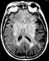
Axial fluid-attenuated inversion recovery MRI image demonstrating tumor-related infiltration involving lenticular nuclei (Arrow).

Axial fluid-attenuated inversion recovery MRI image demonstrating tumor-related infiltration involving both temporal lobes (Short arrow), and the substantia nigra (Long arrow).


See also
References
- ↑ "Fluid-attenuated inversion recovery magnetic resonance imaging detects cortical and juxtacortical multiple sclerosis lesions". Archives of Neurology 58 (5): 742–8. May 2001. doi:10.1001/archneur.58.5.742. PMID 11346369.
- ↑ 2.0 2.1 2.2 "Fluid attenuation inversion recovery | Radiology Reference Article | Radiopaedia.org". http://radiopaedia.org/articles/fluid-attenuation-inversion-recovery. Retrieved 2015-12-03.
Further reading
- MRI from Picture to Proton.. Cambridge University Press. April 2017. pp. 40–42.
- MRI: The Basics (2nd ed.). Philadelphia. p. 272.
 |  |

