Software:Protein Imager: Difference between revisions
From HandWiki
Importwiki (talk | contribs) (import) |
Importwiki (talk | contribs) (import) |
||
| Line 5: | Line 5: | ||
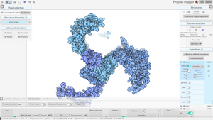
| screenshot alt = Protein Imager interface with example project loaded | | screenshot alt = Protein Imager interface with example project loaded | ||
| caption = Protein Imager interface with example project loaded. | | caption = Protein Imager interface with example project loaded. | ||
| developer = 3D Protein Imaging | | developer = {{URL|https://3dproteinimaging.com|3D Protein Imaging}} | ||
| programming language = Javascript, [[Software:WebGL|WebGL]] | | programming language = Javascript, [[Software:WebGL|WebGL]] | ||
| platform = Web-based. See {{Section link||Browser support}} | | platform = Web-based. See {{Section link||Browser support}} | ||
| Line 13: | Line 13: | ||
| website = {{URL|https://3dproteinimaging.com/protein-imager}} | | website = {{URL|https://3dproteinimaging.com/protein-imager}} | ||
}} | }} | ||
'''Protein Imager'''<ref>{{Cite journal| | '''Protein Imager'''<ref>{{Cite journal |last1=Tomasello |first1=Gianluca |last2=Armenia |first2=Ilaria |last3=Molla |first3=Gianluca |date=2020-05-01 |title=The Protein Imager: a full-featured online molecular viewer interface with server-side HQ-rendering capabilities |url=https://academic.oup.com/bioinformatics/article/36/9/2909/5701652 |journal=Bioinformatics |language=en |volume=36 |issue=9 |pages=2909–2911 |doi=10.1093/bioinformatics/btaa009 |pmid=31930403 |issn=1367-4803}}</ref><ref>{{Cite web |date=2024-02-06 |title=3D Protein Imager a PyMOL/Qutemol web alternative |url=https://bcrf.biochem.wisc.edu/2024/02/06/3d-protein-imager-a-pymol-qutemol-web-alternative/ |access-date=2024-05-02 |website=Biochemistry Computational Research Facility (BCRF) |language=en-US}}</ref>'''<ref>{{Cite web |title=About Protein Imager |url=https://3dproteinimaging.com/about-protein-imager/ |access-date=2023-12-05 |website=3D Protein Imaging |language=en-GB}}</ref>'''<ref>{{Cite web |last=RCSB |first=Protein Data Bank |title=Molecular Graphics Software |url=https://www.rcsb.org/docs/additional-resources/molecular-graphics-software |access-date=2023-12-15 |website=www.rcsb.org |language=en-US}}</ref><ref>{{Cite web |last= |title=Illustrate: Non-photorealistic Biomolecular Illustration |url=https://ccsb.scripps.edu/illustrate/ |access-date=2023-12-15 |website=Illustrate |language=en-US}}</ref><ref>{{Cite web |title=Protein Imager |url=https://my.labbrowser.com/store/app/3-d-protein-imaging |access-date=2024-05-02 |website=my.labbrowser.com}}</ref><ref>{{Cite web |title=Visualizing and Comparing Molecular Structures in Google Colab |url=https://colab.research.google.com/github/pb3lab/ibm3202/blob/master/tutorials/lab02_molviz.ipynb#scrollTo=osOd5k9E03KV |access-date=2024-05-02 |website=colab.research.google.com |language=en}}</ref><ref>{{Cite web |title=«The Protein Imager»: un visualizzatore molecolare dal gruppo di ricerca «The Protein Factory 2.0» {{!}} Università degli studi dell'Insubria |url=https://archivio.uninsubria.it/notizie/%C2%AB-protein-imager%C2%BB-un-visualizzatore-molecolare-dal-gruppo-di-ricerca-%C2%AB-protein-factory-20%C2%BB |access-date=2023-12-15 |website=archivio.uninsubria.it |language=it}}</ref><ref>{{Cite web |last=Noroozi |first=Fereshteh |title=Exploring 3D Protein Imaging Web Service: A Step-by-Step Guide |url=https://www.youtube.com/watch?v=uqL3uSRmS38 |url-status= |website=youtube}}</ref> is a web-based [[Molecular graphics|molecular graphics interface]] being developed by [https://3dproteinimaging.com 3D Protein Imaging] that can be used to visualize [[Biology:Macromolecule|macromolecues]] and obtain publication-quality [[Biology:Molecular modelling|molecular illustrations.]]<ref>{{Cite journal |last1=Langendonk |first1=R. Frèdi |last2=Neill |first2=Daniel R. |last3=Fothergill |first3=Joanne L. |date=2021 |title=The Building Blocks of Antimicrobial Resistance in Pseudomonas aeruginosa: Implications for Current Resistance-Breaking Therapies |journal=Frontiers in Cellular and Infection Microbiology |volume=11 |doi=10.3389/fcimb.2021.665759 |issn=2235-2988 |pmc=8085337 |pmid=33937104 |doi-access=free }}</ref><ref>{{Cite journal |last1=Leitão |first1=Ana Lúcia |last2=Enguita |first2=Francisco J. |date=2022-01-18 |title=A Structural View of miRNA Biogenesis and Function |journal=Non-Coding RNA |language=en |volume=8 |issue=1 |pages=10 |doi=10.3390/ncrna8010010 |issn=2311-553X |pmc=8874510 |pmid=35202084 |doi-access=free }}</ref><ref>{{Cite journal |last1=Ismail |first1=Helene |last2=Shakkour |first2=Zaynab |last3=Tabet |first3=Maha |last4=Abdelhady |first4=Samar |last5=Kobaisi |first5=Abir |last6=Abedi |first6=Reem |last7=Nasrallah |first7=Leila |last8=Pintus |first8=Gianfranco |last9=Al-Dhaheri |first9=Yusra |last10=Mondello |first10=Stefania |last11=El-Khoury |first11=Riyad |last12=Eid |first12=Ali H. |last13=Kobeissy |first13=Firas |last14=Salameh |first14=Johnny |date=2020-10-01 |title=Traumatic Brain Injury: Oxidative Stress and Novel Anti-Oxidants Such as Mitoquinone and Edaravone |journal=Antioxidants |language=en |volume=9 |issue=10 |pages=943 |doi=10.3390/antiox9100943 |issn=2076-3921 |pmc=7601591 |pmid=33019512 |doi-access=free }}</ref><ref>{{Cite journal |last1=Ovejero |first1=Jesus G. |last2=Armenia |first2=Ilaria |last3=Serantes |first3=David |last4=Veintemillas-Verdaguer |first4=Sabino |last5=Zeballos |first5=Nicoll |last6=López-Gallego |first6=Fernando |last7=Grüttner |first7=Cordula |last8=de la Fuente |first8=Jesús M. |last9=Puerto Morales |first9=María del |last10=Grazu |first10=Valeria |date=2021-09-08 |title=Selective Magnetic Nanoheating: Combining Iron Oxide Nanoparticles for Multi-Hot-Spot Induction and Sequential Regulation |journal=Nano Letters |language=en |volume=21 |issue=17 |pages=7213–7220 |doi=10.1021/acs.nanolett.1c02178 |issn=1530-6984 |pmc=8431726 |pmid=34410726|bibcode=2021NanoL..21.7213O }}</ref> | ||
== Browser support == | == Browser support == | ||
Protein Imager supports current versions of [[Software:Firefox|Firefox]], [[Software:Google Chrome|Chrome]], [[Software:Safari (web browser)|Safari]], and [[Software:Microsoft Edge|Edge]] | Protein Imager supports current versions of [[Software:Firefox|Firefox]], [[Software:Google Chrome|Chrome]], [[Software:Safari (web browser)|Safari]], and [[Software:Microsoft Edge|Edge]], including their respective mobile versions. | ||
== Gallery == | == Gallery == | ||
<gallery> | <gallery> | ||
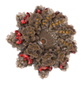
File:Protein Imager high quality illustration example 1 HIV capsid.png|alt=Protein Imager Rendering Example HIV Viral capsid|Protein Imager | File:Protein Imager high quality illustration example 1 HIV capsid.png|alt=Protein Imager Rendering Example HIV Viral capsid|Protein Imager rendering Example. HIV Viral capsid | ||
File:Protein Imager high quality illustration example 2 parathyroid hormone 1 receptor.png|alt=Protein Imager Rendering Example Membrane protein|Protein Imager | File:Protein Imager high quality illustration example 2 parathyroid hormone 1 receptor.png|alt=Protein Imager Rendering Example Membrane protein|Protein Imager rendering Example. Membrane protein | ||
File:Protein Imager molecular rendering example 3 CsgFG complex.png|alt=Protein Imager Rendering Example Protein complex|Protein Imager | File:Protein Imager molecular rendering example 3 CsgFG complex.png|alt=Protein Imager Rendering Example Protein complex|Protein Imager rendering Example. Protein complex | ||
</gallery> | </gallery> | ||
Latest revision as of 17:43, 3 May 2024
 Protein Imager interface with example project loaded. | |
| Developer(s) | 3D Protein Imaging |
|---|---|
| Written in | Javascript, WebGL |
| Platform | Web-based. See § Browser support |
| Available in | English |
| Type | Molecular graphics |
| License | Proprietary |
| Website | 3dproteinimaging |
Protein Imager[1][2][3][4][5][6][7][8][9] is a web-based molecular graphics interface being developed by 3D Protein Imaging that can be used to visualize macromolecues and obtain publication-quality molecular illustrations.[10][11][12][13]
Browser support
Protein Imager supports current versions of Firefox, Chrome, Safari, and Edge, including their respective mobile versions.
Gallery
References
- ↑ Tomasello, Gianluca; Armenia, Ilaria; Molla, Gianluca (2020-05-01). "The Protein Imager: a full-featured online molecular viewer interface with server-side HQ-rendering capabilities" (in en). Bioinformatics 36 (9): 2909–2911. doi:10.1093/bioinformatics/btaa009. ISSN 1367-4803. PMID 31930403. https://academic.oup.com/bioinformatics/article/36/9/2909/5701652.
- ↑ "3D Protein Imager a PyMOL/Qutemol web alternative" (in en-US). 2024-02-06. https://bcrf.biochem.wisc.edu/2024/02/06/3d-protein-imager-a-pymol-qutemol-web-alternative/.
- ↑ "About Protein Imager" (in en-GB). https://3dproteinimaging.com/about-protein-imager/.
- ↑ RCSB, Protein Data Bank. "Molecular Graphics Software" (in en-US). https://www.rcsb.org/docs/additional-resources/molecular-graphics-software.
- ↑ "Illustrate: Non-photorealistic Biomolecular Illustration" (in en-US). https://ccsb.scripps.edu/illustrate/.
- ↑ "Protein Imager". https://my.labbrowser.com/store/app/3-d-protein-imaging.
- ↑ "Visualizing and Comparing Molecular Structures in Google Colab" (in en). https://colab.research.google.com/github/pb3lab/ibm3202/blob/master/tutorials/lab02_molviz.ipynb#scrollTo=osOd5k9E03KV.
- ↑ "«The Protein Imager»: un visualizzatore molecolare dal gruppo di ricerca «The Protein Factory 2.0» | Università degli studi dell'Insubria" (in it). https://archivio.uninsubria.it/notizie/%C2%AB-protein-imager%C2%BB-un-visualizzatore-molecolare-dal-gruppo-di-ricerca-%C2%AB-protein-factory-20%C2%BB.
- ↑ Noroozi, Fereshteh. "Exploring 3D Protein Imaging Web Service: A Step-by-Step Guide". https://www.youtube.com/watch?v=uqL3uSRmS38.
- ↑ Langendonk, R. Frèdi; Neill, Daniel R.; Fothergill, Joanne L. (2021). "The Building Blocks of Antimicrobial Resistance in Pseudomonas aeruginosa: Implications for Current Resistance-Breaking Therapies". Frontiers in Cellular and Infection Microbiology 11. doi:10.3389/fcimb.2021.665759. ISSN 2235-2988. PMID 33937104.
- ↑ Leitão, Ana Lúcia; Enguita, Francisco J. (2022-01-18). "A Structural View of miRNA Biogenesis and Function" (in en). Non-Coding RNA 8 (1): 10. doi:10.3390/ncrna8010010. ISSN 2311-553X. PMID 35202084.
- ↑ Ismail, Helene; Shakkour, Zaynab; Tabet, Maha; Abdelhady, Samar; Kobaisi, Abir; Abedi, Reem; Nasrallah, Leila; Pintus, Gianfranco et al. (2020-10-01). "Traumatic Brain Injury: Oxidative Stress and Novel Anti-Oxidants Such as Mitoquinone and Edaravone" (in en). Antioxidants 9 (10): 943. doi:10.3390/antiox9100943. ISSN 2076-3921. PMID 33019512.
- ↑ Ovejero, Jesus G.; Armenia, Ilaria; Serantes, David; Veintemillas-Verdaguer, Sabino; Zeballos, Nicoll; López-Gallego, Fernando; Grüttner, Cordula; de la Fuente, Jesús M. et al. (2021-09-08). "Selective Magnetic Nanoheating: Combining Iron Oxide Nanoparticles for Multi-Hot-Spot Induction and Sequential Regulation" (in en). Nano Letters 21 (17): 7213–7220. doi:10.1021/acs.nanolett.1c02178. ISSN 1530-6984. PMID 34410726. Bibcode: 2021NanoL..21.7213O.
 |


