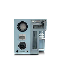
Engineering:Celloscope automated cell counter
Celloscope automated cell counter was developed in the 50s for enumeration of erythrocytes, leukocytes, and thrombocytes in blood samples.[1] Together with the Coulter counter, the Celloscope analyzer can be considered one of the predecessors of today’s automated hematology analyzers, as the principle of the electrical impedance method is still utilized in cell counters installed in clinical laboratories around the world.[2][3]
History
The Celloscope was developed for the Swedish company AB Lars Ljungberg & Co under the direction of engineer Erik Öhlin at Linson Instrument AB.[4] In an interview published in the Clinical Biochemistry in the Nordics, a membership magazine for the Nordic Association for Clinical Chemistry, Lars Ljungberg explains that he and his coworkers had been considering different solutions for counting blood cells for some time when they came across a method presented by the American Navy on how particles could be counted when allowed to pass a capillary hole through which a weak direct current was passed simultaneously.[5][6] The Celloscope method exploits the feature of blood cells not being conductive and therefore make interruptions (pulses) to the current, which then can be counted.[7] What Ljungberg and coworkers did not know was that Wallace H. Coulter in Chicago had applied for and received a patent on the particle count principle in 1953.[8] When presented at a German tradeshow in September 1957, the Celloscope counter was examined by Dr. George Brecher, the first author of one of the NIH evaluations of the Coulter counter.[9] In a letter to Coulter, Brecher reported about what he thought was a close functional copy of the Coulter counter, yet with simpler electronics and an integrated sample stand, creating a both smaller and less costly instrument for use in clinical applications.[9] When the Celloscope was introduced to the market in the early 60s, a lawsuit was filed by Coulter Electronics Inc against AB Lars Ljungberg & Co for alleged infringement of the American patent.[10] After many and long negotiations, the companies came to the agreement to compensate Coulter for the sales that had been made in USA and some European countries where he had the patent and that AB Lars Ljungberg & Co was free to sell their analyzer in other regions.[9][6]
Method principle
Before the introduction of the first automated cell counters, hematologists were referred to manual cell count under the microscope.[2][11] The Celloscope method for automated counting of blood cells was described in an article by Öhlin in 1958.[1][12] In the described method, cells in a saline (conductive) solution are allowed to pass through a capillary with a length and diameter corresponding to the size of blood cells. At the same time, an electric current passes the capillary, and each cell then gives rise to an electric pulse through the increase in resistance that it causes in the electric circuit. The number of pulses is recorded and corresponds to the number of cells in a certain volume. Diluting the blood sample to a sufficient extent for the distance between the cells when passing through the capillary to be greater than the dimension of the cells and capillary ensures that each cell is counted individually. As cells are counted in an absolute volume of the suspension, the number of cells in mm3 of whole blood can be calculated using the dilution factor. The described automated Celloscope cell count method enabled an improved accuracy compared with manual examination by microscopy, while decreasing manual work for the operator. The method allows 50 000 cells to be counted in about 45 seconds, with high accuracy. The Celloscope counter was also equipped with a discriminator, or electrical threshold, which allows only pulses above a certain size to be counted, enabling different blood cells to be counted. For example, set to a threshold of 3 µm, all cells are counted. A re-count at a threshold of 4 µm allows calculation of the number of cells of a size between 3 and 4 µm from the total counts. Diluting the blood sample 1/80 000 in a physiological saline solution allows enumeration of the erythrocytes, as the number of leukocytes does not affect the result more than by about 1/1 000 000. For the leukocyte count, cells in the same sample are hemolyzed with saponin or cetrimide so that only the nuclei of the leukocytes are counted.[6][13][14] For platelet count, a smaller capillary diameter is used. To identify cell morphologies and variants that the counter cannot detect, microscopy remains an essential complement to the automated cell count method.[15]
Successor cell counters
The impedance method used in the Celloscope analyzer has been further developed to allow counting also of leukocyte subgroups.[2][3] In addition to the cell counts, modern hematology analyzers are also capable of reporting parameters related to cell size, hemoglobin concentration, as well as a range of calculated parameters, for a complete blood count (CBC). These analyzers were initially intended to be used in hospital laboratories, as they required a skilled staff and a high sample load to justify their relatively high cost, however, with the increasing need for decentralized healthcare, the demand for simpler analyzers emerged and prompted the development of benchtop cell counters that could be used in a near-patient clinical setting with a minimum of training.[2][16][17] In 1969, Erik Öhlin founded Swelab Instrument AB (today, Boule Medical AB[18]), and later, the Swelab AutoCounter AC-series was launched to meet the needs of the smaller clinical laboratories.
The cell counters of that time used LED screens for result review. In 1982, Medonic AB, another Swedish company with focus on hematology, was founded.[19] The founders, Ingemar Berndtsson and Abraham Bottema, both had a long history and experience in hematology, clinical chemistry, and blood banking engineering.[20][21][22] In 1985, Medonic AB launched the Cellanalyzer CA 480 system, its first own-developed cell counter with a built-in display that also showed the cell histograms.[23] When computers began to be incorporated into the analyzers, other brands, like the Swelab analyzers, also came with a display. Both targeting the smaller clinical laboratories, Swelab Instrument AB and Medonic AB were competitors on the decentralized hematology testing market.[24][25][26] In the late 90s, both Swelab Instrument AB and Medonic AB were acquired by Boule Diagnostics AB.[27][28] The company has kept the parallel brands and the analyzers are still manufactured from its facilities in Stockholm, Sweden and supplied under the Swelab and Medonic trademarks for the decentralized hematology testing market.[29][30][31][32][33][34]
When Coulter was acquired by Beckman, former Coulter employees Dr. Harold R Crews, Andrew C Swanson, and Donald Grantham founded Clinical Diagnostic Solutions, Inc. (CDS) in 1997, focusing on the development and production of generic reagents and control material. In 2004, CDS was acquired by Boule. By this acquisition, Boule came to master the skills of the development and production of both instruments and the consumables included in a complete hematology system.
References
- ↑ Jump up to: 1.0 1.1 Öhlin, E (1958). "Automatisk cellräknings-metod – speciellt för blodkroppar". Nordisk Medicin 59: 577–578.
- ↑ Jump up to: 2.0 2.1 2.2 2.3 Green, R. (2015). "Development, History, and Future of Automated Cell Counters". Clinics in Laboratory Medicine 35 (1): 1–10. doi:10.1016/j.cll.2014.11.003. PMID 25676368. https://www.sciencedirect.com/science/article/abs/pii/S0272271214001139.
- ↑ Jump up to: 3.0 3.1 Bruegel, M (2015). "Comparison of five automated hematology analyzers in a university hospital setting: Abbott Cell-Dyn Sapphire, Beckman Coulter DxH 800, Siemens Advia 2120i, Sysmex XE-5000, and Sysmex XN-2000". Clin Chem Lab Med 53 (7): 1057–1071. doi:10.1515/cclm-2014-0945. PMID 25720071. https://www.researchgate.net/publication/272836354. Retrieved 12 July 2023.
- ↑ "Telub-Nytt". 14 Jan 1970. https://arkiv.telub.se/pdf/Telub-nytt/1970/01/pdf.pdf.
- ↑ Robinson, J.P. (2013). "Wallace H. Coulter: Decades of invention and discovery". Cytometry 83A (5): 424–438. doi:10.1002/cyto.a.22296. PMID 23596093.
- ↑ Jump up to: 6.0 6.1 6.2 Hagve, T-A (2007). "Fra mikroskop til flowcelle.". Klinisk Biokemi I Norden 19 (2): 8–17. https://www.nfkk.org/wp-content/uploads/kbn-2007-2.pdf. Retrieved 3 March 2023.
- ↑ Cole, K.S. (1928). "Electric impedance of suspensions of spheres". J Gen Physiol 12 (1): 29–36. doi:10.1085/jgp.12.1.29. PMID 19872446.
- ↑ Coulter, W.H. (20 Oct 1953). "Means for counting particles suspended in a fluid". Patent US2656508A. https://portal.unifiedpatents.com/patents/patent/US-2656508-A. Retrieved 3 March 2023.
- ↑ Jump up to: 9.0 9.1 9.2 Marshall, G (2020). The coulter principle: for the good of humankind. Theses and Dissertations-History. doi:10.13023/etd.2020.495. https://doi.org/10.13023/etd.2020.495. Retrieved 3 March 2023.
- ↑ Singer, H.J. (29 Oct 2011). Coulter Electronics Inc. v. A.B. Lars Ljungberg & Co. Supreme Court Transcript of Record with Supporting Pleadings. Gale. U.S. Supreme Court Records. ISBN 978-1270529194.
- ↑ Cadena-Herrera, D. (2015). "Validation of three viable-cell counting methods: Manual, semi-automated, and automated". Biotechnology Reports 7: 9–16. doi:10.1016/j.btre.2015.04.004. PMID 28626709.
- ↑ "CELLOSCOPE Trademark - Registration Number 0850186 - Serial Number 72243164 :: Justia Trademarks" (in en). http://trademarks.justia.com/722/43/celloscope-72243164.html.
- ↑ Lappin, T. R. J. (1972). "An evaluation of the Celloscope 401 electronic blood cell counter". J. Clin. Path. 25 (6): 539–542. doi:10.1136/jcp.25.6.539. PMID 4625438. PMC 477376. https://jcp.bmj.com/content/25/6/539.long.
- ↑ Kvarstein, B (1967). "The Use of an Electronic Particle Counter (Celloscope 101) for Counting Leucocytes". Scandinavian Journal of Clinical and Laboratory Investigation 19 (2): 196–202. doi:10.3109/00365516709093502. https://www.tandfonline.com/doi/abs/10.3109/00365516709093502.
- ↑ Gulati, G (2013). "Purpose and Criteria for Blood Smear Scan, Blood Smear Examination, and Blood Smear Review". Ann Lab Med 33 (1): 1–7. doi:10.3343/alm.2013.33.1.1. PMID 23301216.
- ↑ Kallner, A. (May 2019). Från hantverk till industri. Om utvecklingen av klinisk kemi på Karolinska sjukhuset och i SLL. http://www.wikiks.se/wp-content/uploads/2019/05/19-05-12-Klinisk-kemi-p%C3%A5-KSbw2.pdf. Retrieved 3 March 2023.
- ↑ Mooney, C (2019). "Point of care testing in general haematology". BJH 187 (3): 265–403. doi:10.1111/bjh.16208. PMID 31578729.
- ↑ "Boule Medical AB". https://foretagsinfo.bolagsverket.se/sok-foretagsinformation-web/foretag/5561286542/foretagsform/AB.
- ↑ "Medonic AB". https://foretagsinfo.bolagsverket.se/sok-foretagsinformation-web/foretag/5562145093/foretagsform/AB.
- ↑ Berndtsson, I. "SE456866B". European Patent Office. https://worldwide.espacenet.com/patent/search/family/020369807/publication/SE456866B?q=SE456866B.
- ↑ Berndtsson, I. "EP0311588A2". European Patent Office. https://worldwide.espacenet.com/patent/search/family/020369875/publication/EP0311588A2?q=EP0311588A2.
- ↑ Berndtsson, I. "SE507956C2". European Patent Office. https://worldwide.espacenet.com/patent/search/family/020404686/publication/SE507956C2?q=SE507956C2.
- ↑ Vonderschmitt, D.J. (2011). Laboratory Organization. Automation. Walter de Gruyter. ISBN 978-3110888454.
- ↑ Ige, O.M. (2012). "Atopy is a risk factor for adult asthma in urban community of Southwestern Nigeria". Lung India 29 (2): 114–119. doi:10.4103/0970-2113.95301. PMID 22628923.
- ↑ Ponampalam, P. (2012). "Comparison of Full Blood Count Parameters Using Capillary and Venous Samples in Patients Presenting to the Emergency Department". ISRN Emergency Medicine 2012: 1–6. doi:10.5402/2012/508649. https://downloads.hindawi.com/archive/2012/508649.pdf.
- ↑ Wallen, Nh; Larsson, Pt; Broijersen, A.; Andersson, A.; Hjemdahl, P. (1993). "Effects of an oral dose of isosorbide dinitrate on platelet function and fibrinolysis in healthy volunteers." (in en). British Journal of Clinical Pharmacology 35 (2): 143–151. doi:10.1111/j.1365-2125.1993.tb05680.x. PMID 8443032.
- ↑ Ben Hayun, H. (2020). Optimization of production-flow for hematology instruments at Boule Medical AB. Thesis KTH. https://www.diva-portal.org/smash/get/diva2:1465220/FULLTEXT01.pdf. Retrieved 12 July 2023.
- ↑ "Boule Diagnostics AB (publ) Year-End Report 2016". 2016. http://mb.cision.com/Main/272/2243118/659968.pdf.
- ↑ "Patent-och registreringsverket". https://www.prv.se/.
- ↑ "Patent-och registreringsverket". https://www.prv.se/.
- ↑ Muhammad, N. (2020). "Thrombocytopenia in patients diagnosed with Malaria in District Buner, KP". JRMC 24 (4): 316–321. doi:10.37939/JRMC.V24I4.1321.
- ↑ Muresan, E-M. (2022). "Admission Emergency Department Point-of-care Biomarkers for Prediction of Early Mortality in Spontaneous Intracerebral Hemorrhage". In Vivo 36 (3): 1534–1543. doi:10.21873/invivo.12864. PMID 35478162. PMC 9087056. https://iv.iiarjournals.org/content/invivo/36/3/1534.full.pdf.
- ↑ Ali, Hussein Noori; Ali, Kameran Mohammed; Rostam, Hassan Muhammad; Ali, Ayad M.; Tawfeeq, Hassan Mohammad; Fatah, Mohammed Hassan; Figueredo, Grazziela P. (2022-08-01). "Clinical laboratory parameters and comorbidities associated with severity of coronavirus disease 2019 (COVID-19) in Kurdistan Region of Iraq" (in en). Practical Laboratory Medicine 31: e00294. doi:10.1016/j.plabm.2022.e00294. ISSN 2352-5517. PMID 35873658.
- ↑ Latif, M. (2022). "Frequency and Severity of Anemia in Pregnant Women at Gwadar Development Authority Hospital, Gwadar, Balochistan". Pak Armed Forces Med J 72 (Suppl-2): S204. https://www.researchgate.net/publication/361151658.
External links
 |


