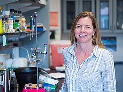
Biography:Gia Voeltz
Gia Voeltz | |
|---|---|
 | |
| Born | Gia Voeltz Bloomington, Indiana, United States |
| Alma mater | University of California Santa Cruz (BS) Yale University (PhD) Harvard Medical School (Postdoctoral) |
| Known for | discovering the function of the Reticulon protein family |
| Awards | Member: National Academy of Sciences 2023 Fellow: American Society for Cell Biology 2023 Investigator: Howard Hughes Medical Institute 2018 Scholar: Howard Hughes Medical Institute 2016 |
| Scientific career | |
| Fields | |
| Institutions | |
| Thesis | mRNA Stability is Regulated during Early Development by AU-rich Sequences and a Novel Poly(A) Binding Protein, ePAB (2001) |
| Doctoral advisor | Joan A. Steitz |
Gia Voeltz is an American cell biologist. She is a professor of Molecular, Cellular and Developmental Biology at the University of Colorado Boulder and a Howard Hughes Medical Institute Investigator. She is known for her research identifying the factors and unraveling the mechanisms that determine the structure and dynamics of the largest organelle in the cell: the endoplasmic reticulum. [1] [2] Her lab has produced paradigm shifting studies on organelle membrane contact sites that have revealed that most cytoplasmic organelles are not isolated entities but are instead physically tethered to an interconnected ER membrane network. [3] [4] [5]
Her research has revealed the fundamental nature of these ER contact sites in regulating the biogenesis of other organelles at positions where they are tethered and closely opposed. [6] [7] [8] [9]
Early life and education
Gia Voeltz grew up in several different states including Indiana , Hawaii, Minnesota and Upstate New York, where she graduated from Chenango Forks High School. She attended university at the University of California Santa Cruz where she majored in Biochemistry and Molecular Biology. She performed her senior thesis work in the lab of Manny Ares [10] on pre-spliceosome assembly in yeast. [11] This experience in the Ares lab at UC Santa Cruz inspired her to become a scientist. Her early undergraduate research studying RNA processing led her to pursue a PhD thesis in the Department of Molecular Biophysics and Biochemistry at Yale University in the lab of Joan A. Steitz, a leading figure in RNA biology. Her PhD research investigated how mRNA stability was regulated during different stages of early development using Xenopus eggs and extract as a model system. [12] [13]
She then moved to Harvard Medical School to join the lab of Tom Rapoport as a Jane Coffin Childs postdoctoral fellow.
Career
Gia Voeltz was trained as an RNA biologist but made a major switch in scientific sub-fields when she moved to Tom Rapoport’s lab as a postdoc to study how organelles get their shape. As a postdoc, she set out to identify how membrane proteins generate the elaborate shape of the ER. To do this, she used biochemical fractionation of a Xenopus egg in vitro assay for ER network formation.[14] Her postdoctoral studies identified the Reticulon family of ER membrane proteins and demonstrated their conserved role in generating the structure of the tubular ER network.[2] The hairpin "wedge" mechanism proposed was that Reticulon has two short hairpin transmembrane domains that occupy more area in the outer leaflet to generate the high membrane curvature found in tubules.[2]
Gia Voeltz moved to University of Colorado Boulder in 2006[15] to start her own lab. Her lab leveraged spinning disk confocal microscopy to visualize the reticulon-generated dynamic tubular ER network in live cells at high resolution.[3] This led to the observation that ER tubule dynamics often occurred at positions where the ER tubules were tightly tethered to other dynamic organelles like endosomes and mitochondria.[3][4]
Multi-color live cell fluorescence imaging complemented by high resolution electron microscopy and tomography revealed that the vast majority of endosomes and mitochondria are tethered to the ER at contact sites. In a hallmark paper published in 2011, Voeltz lab, in a collaboration with Jodi Nunnari’s lab, showed that ER tubules wrap around mitochondria to define the position where mitochondria constrict and divide in animal and yeast cells.[6]
Her lab has gone on to show that ER contact sites also regulate early and late endosome fission,[7][8] RNA granule division,[9] and mitochondrial fusion.[16] [17] These works establish the ER network as a master regulator of organelle biogenesis through ER contact sites.[5][18]
Gia Voeltz became a Howard Hughes Medical Institute Scholar in 2016[19] and a Howard Hughes Medical Institute Investigator in 2018.[20][21] She was elected to the National Academy of Sciences in 2023.[22]
Awards and honors
- Elected a Member of the National Academy of Sciences (2023)[22][23]
- Elected a Fellow of the American Society of Cell Biology (2023)[24]
- Appointed an Investigator of the Howard Hughes Medical Institute (2018)[21]
- Howard Hughes Medical Institute Scholar Award (2016)[19]
- Porter Lecture Award from the American Society of Cell Biology (2015)
- American Cancer Society Research Scholar Award (2013)
- Provost's Faculty Achievement Award, University of Colorado Boulder (2012)
- Günter Blobel Early Career Award (2012)[25]
- Searle Scholars Early Career Award (2007)
- Jane Coffin Childs Postdoctoral Fellowship Award (2001)
- Graduated with High Honors, University of California Santa Cruz (1994)
Institutions
- University of Colorado Boulder 2006–present
- Howard Hughes Medical Institute 2018–present
- PhD Advisor: Joan A. Steitz, Yale University, 1995–2001
- Postdoc Advisor: Tom Rapoport, Harvard Medical School 2001–2006
References
- ↑ "Form follows function: the importance of endoplasmic reticulum shape". Annual Review of Biochemistry 84: 791–811. January 2015. doi:10.1146/annurev-biochem-072711-163501. PMID 25580528.
- ↑ 2.0 2.1 2.2 "A class of membrane proteins shaping the tubular endoplasmic reticulum". Cell 124 (3): 573–586. 10 February 2006. doi:10.1016/j.cell.2005.11.047. PMID 16469703.
- ↑ 3.0 3.1 3.2 "ER sliding dynamics and ER-mitochondrial contacts occur on acetylated microtubules". Journal of Cell Biology 190 (3): 363–375. 9 August 2010. doi:10.1083/jcb.200911024. PMID 20696706.
- ↑ 4.0 4.1 "Endoplasmic reticulum-endosome contact increases as endosomes traffic and mature". Molecular Biology of the Cell 24 (7): 1030–1040. 6 February 2013. doi:10.1091/mbc.E12-10-0733. PMID 23389631.
- ↑ 5.0 5.1 "Here, there, and everywhere: The importance of ER membrane contact sites". Science 361 (6401). 3 August 2018. doi:10.1126/science.aan5835. PMID 30072511.
- ↑ 6.0 6.1 "ER tubules mark sites of mitochondrial division". Science 334 (6954): 358–362. 21 October 2011. doi:10.1126/science.1207385. PMID 21885730. Bibcode: 2011Sci...334..358F.
- ↑ 7.0 7.1 "ER contact sites define the position and timing of endosome fission". Cell 159 (5): 1027–1041. 20 November 2014. doi:10.1016/j.cell.2014.10.023. PMID 25416943.
- ↑ 8.0 8.1 "A Novel Class of ER Membrane Proteins Regulates ER-Associated Endosome Fission". Cell 175 (1): 254–265. 13 September 2018. doi:10.1016/j.cell.2018.08.030. PMID 30220460.
- ↑ 9.0 9.1 "Endoplasmic reticulum contact sites regulate the dynamics of membraneless organelles". Science 367 (6477). 31 January 2020. doi:10.1126/science.aay7108. PMID 32001628.
- ↑ "Dr. Manuel Ares". University of California, Santa Cruz. https://mcd.ucsc.edu/faculty/ares.html.
- ↑ "ATP requirement for Prp5p function is determined by Cus2p and the structure of U2 small nuclear RNA". Proceedings of the National Academy of Sciences of the United States of America 100 (24): 13857–13862. 10 November 2003. doi:10.1073/pnas.2036312100. PMID 14610285. Bibcode: 2003PNAS..10013857P.
- ↑ "AUUUA Sequences Direct mRNA Deadenylation Uncoupled from Decay during Xenopus Early Development". Molecular and Cellular Biology 18 (12): 7537–7545. 23 August 1998. doi:10.1128/MCB.18.12.7537. PMID 9819439.
- ↑ "A novel embryonic poly(A) binding protein, ePAB, regulates mRNA deadenylation in Xenopus egg extracts". Genes and Development 15 (6): 774–788. 15 March 2001. doi:10.1101/gad.872201. PMID 11274061.
- ↑ "In vitro formation of the endoplasmic reticulum occurs independently of microtubules by a controlled fusion reaction". Journal of Cell Biology 148 (5): 883–898. 6 March 2000. doi:10.1083/jcb.148.5.883. PMID 10704440.
- ↑ "Building a path in cell biology". Molecular Biology of the Cell 23 (21): 4145–4147. 13 October 2017. doi:10.1091/mbc.E12-05-0382. PMID 23112222.
- ↑ "Fission and fusion machineries converge at ER contact sites to regulate mitochondrial morphology". Journal of Cell Biology 219 (4). 25 February 2020. doi:10.1083/jcb.201911122. PMID 32328629.
- ↑ "An ER phospholipid hydrolase drives ER-associated mitochondrial constriction for fission and fusion". eLife 11. 30 Nov 2022. doi:10.7554/eLife.84279. PMID 36448541.
- ↑ Voeltz, Gia (speaker) (22 May 2019). Factors and Functions of Organelle Membrane Contact Sites. iBiology. Retrieved 18 January 2024 – via Youtube.
- ↑ 19.0 19.1 "HHMI 2016 Faculty Scholars". HHMI. https://media.hhmi.org/FacultyScholars2016-gallery/.
- ↑ "HHMI Bets Big On 19 New Investigators". HHMI. 23 May 2018. https://www.hhmi.org/news/hhmi-bets-big-19-new-investigators.
- ↑ 21.0 21.1 "Cellular cartographer Voeltz named HHMI investigator, granted $8 million". University of Colorado Boulder. 23 May 2018. https://www.colorado.edu/today/2018/05/23/cellular-cartographer-voeltz-named-hhmi-investigator-granted-8-million.
- ↑ 22.0 22.1 "Pioneering biologist elected to National Academy of Sciences". University of Colorado Boulder. 12 May 2023. https://www.colorado.edu/asmagazine/2023/05/12/pioneering-biologist-elected-national-academy-sciences.
- ↑ "Gia K. Voeltz". National Academy of Sciences. https://www.nasonline.org/member-directory/members/20056796.html.
- ↑ "Nineteen distinguished scientists recognized as 2023 ASCB Fellows". American Society for Cell Biology. 2 August 2023. https://www.ascb.org/society-news/nineteen-distinguished-scientists-recognized-as-2023-ascb-fellows/.
- ↑ "Günter Blobel Early Career Award". American Society for Cell Biology. 2012. https://www.ascb.org/award/early-career-life-scientist-award/.
 |

