Biology:Corpus cavernosum of clitoris
| Corpus cavernosum of clitoris | |
|---|---|
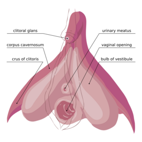
 The internal anatomy of the human vulva, with the clitoral hood and labia minora indicated as lines. | |
| Details | |
| Identifiers | |
| Latin | corpus cavernosum clitoridis |
| Anatomical terminology | |
The corpus cavernosum of clitoris is one of a pair of sponge-like regions of erectile tissue which contain most of the blood in the clitoris during clitoral erection. This is homologous to the corpus cavernosum penis in the male; the body of the clitoris contains erectile tissue in a pair of corpora cavernosa (literally "cave-like bodies"), with a recognizably similar structure.
Anatomy
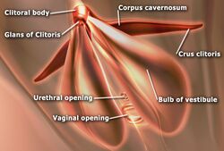
The two corpora cavernosa are expandable erectile tissues which fill with blood during clitoral erection. These formations are made of a sponge-like tissue containing irregular blood-filled spaces lined by endothelium and separated by connective tissue septa. United together along their medial surfaces by an incomplete fibrous pectiniform septum; each corpus is connected to the rami of the pubis and ischium by a clitoral crus.[1]
The female anatomy has two vestibular bulbs beneath the skin of the labia minora (at the entrance to the vagina), which expand at the same time as the glans clitoridis to cap the ends of the corpora cavernosa. This is homologous with the corpus spongiosum of the male anatomy.
Physiology
In some circumstances, release of nitric oxide precedes relaxation of the clitoral cavernosal artery and nearby muscle, in a process similar to male arousal. More blood flows in through the clitoral cavernosal artery, the pressure in the corpora cavernosa clitoridis rises, and the clitoris is engorged with blood. This leads to extrusion of the glans clitoridis and enhanced sensitivity to physical contact.
References
- ↑ Gray, Henry (1918). Atlas of the Human Body. Lea & Febiger.
External links


