Software:Protein Imager: Difference between revisions
From HandWiki
Importwiki (talk | contribs) (import) |
Importwiki (talk | contribs) (import) |
||
| Line 5: | Line 5: | ||
| screenshot alt = Protein Imager interface with example project loaded | | screenshot alt = Protein Imager interface with example project loaded | ||
| caption = Protein Imager interface with example project loaded. | | caption = Protein Imager interface with example project loaded. | ||
| developer = 3D Protein Imaging | | developer = {{URL|https://3dproteinimaging.com|3D Protein Imaging}} | ||
| programming language = Javascript, [[Software:WebGL|WebGL]] | | programming language = Javascript, [[Software:WebGL|WebGL]] | ||
| platform = Web-based. See {{Section link||Browser support}} | | platform = Web-based. See {{Section link||Browser support}} | ||
| Line 13: | Line 13: | ||
| website = {{URL|https://3dproteinimaging.com/protein-imager}} | | website = {{URL|https://3dproteinimaging.com/protein-imager}} | ||
}} | }} | ||
'''Protein Imager'''<ref>{{Cite journal| | '''Protein Imager'''<ref>{{Cite journal |last1=Tomasello |first1=Gianluca |last2=Armenia |first2=Ilaria |last3=Molla |first3=Gianluca |date=2020-05-01 |title=The Protein Imager: a full-featured online molecular viewer interface with server-side HQ-rendering capabilities |url=https://academic.oup.com/bioinformatics/article/36/9/2909/5701652 |journal=Bioinformatics |language=en |volume=36 |issue=9 |pages=2909–2911 |doi=10.1093/bioinformatics/btaa009 |pmid=31930403 |issn=1367-4803}}</ref><ref>{{Cite web |date=2024-02-06 |title=3D Protein Imager a PyMOL/Qutemol web alternative |url=https://bcrf.biochem.wisc.edu/2024/02/06/3d-protein-imager-a-pymol-qutemol-web-alternative/ |access-date=2024-05-02 |website=Biochemistry Computational Research Facility (BCRF) |language=en-US}}</ref>'''<ref>{{Cite web |title=About Protein Imager |url=https://3dproteinimaging.com/about-protein-imager/ |access-date=2023-12-05 |website=3D Protein Imaging |language=en-GB}}</ref>'''<ref>{{Cite web |last=RCSB |first=Protein Data Bank |title=Molecular Graphics Software |url=https://www.rcsb.org/docs/additional-resources/molecular-graphics-software |access-date=2023-12-15 |website=www.rcsb.org |language=en-US}}</ref><ref>{{Cite web |last= |title=Illustrate: Non-photorealistic Biomolecular Illustration |url=https://ccsb.scripps.edu/illustrate/ |access-date=2023-12-15 |website=Illustrate |language=en-US}}</ref><ref>{{Cite web |title=Protein Imager |url=https://my.labbrowser.com/store/app/3-d-protein-imaging |access-date=2024-05-02 |website=my.labbrowser.com}}</ref><ref>{{Cite web |title=Visualizing and Comparing Molecular Structures in Google Colab |url=https://colab.research.google.com/github/pb3lab/ibm3202/blob/master/tutorials/lab02_molviz.ipynb#scrollTo=osOd5k9E03KV |access-date=2024-05-02 |website=colab.research.google.com |language=en}}</ref><ref>{{Cite web |title=«The Protein Imager»: un visualizzatore molecolare dal gruppo di ricerca «The Protein Factory 2.0» {{!}} Università degli studi dell'Insubria |url=https://archivio.uninsubria.it/notizie/%C2%AB-protein-imager%C2%BB-un-visualizzatore-molecolare-dal-gruppo-di-ricerca-%C2%AB-protein-factory-20%C2%BB |access-date=2023-12-15 |website=archivio.uninsubria.it |language=it}}</ref><ref>{{Cite web |last=Noroozi |first=Fereshteh |title=Exploring 3D Protein Imaging Web Service: A Step-by-Step Guide |url=https://www.youtube.com/watch?v=uqL3uSRmS38 |url-status= |website=youtube}}</ref> is a web-based [[Molecular graphics|molecular graphics interface]] being developed by [https://3dproteinimaging.com 3D Protein Imaging] that can be used to visualize [[Biology:Macromolecule|macromolecues]] and obtain publication-quality [[Biology:Molecular modelling|molecular illustrations.]]<ref>{{Cite journal |last1=Langendonk |first1=R. Frèdi |last2=Neill |first2=Daniel R. |last3=Fothergill |first3=Joanne L. |date=2021 |title=The Building Blocks of Antimicrobial Resistance in Pseudomonas aeruginosa: Implications for Current Resistance-Breaking Therapies |journal=Frontiers in Cellular and Infection Microbiology |volume=11 |doi=10.3389/fcimb.2021.665759 |issn=2235-2988 |pmc=8085337 |pmid=33937104 |doi-access=free }}</ref><ref>{{Cite journal |last1=Leitão |first1=Ana Lúcia |last2=Enguita |first2=Francisco J. |date=2022-01-18 |title=A Structural View of miRNA Biogenesis and Function |journal=Non-Coding RNA |language=en |volume=8 |issue=1 |pages=10 |doi=10.3390/ncrna8010010 |issn=2311-553X |pmc=8874510 |pmid=35202084 |doi-access=free }}</ref><ref>{{Cite journal |last1=Ismail |first1=Helene |last2=Shakkour |first2=Zaynab |last3=Tabet |first3=Maha |last4=Abdelhady |first4=Samar |last5=Kobaisi |first5=Abir |last6=Abedi |first6=Reem |last7=Nasrallah |first7=Leila |last8=Pintus |first8=Gianfranco |last9=Al-Dhaheri |first9=Yusra |last10=Mondello |first10=Stefania |last11=El-Khoury |first11=Riyad |last12=Eid |first12=Ali H. |last13=Kobeissy |first13=Firas |last14=Salameh |first14=Johnny |date=2020-10-01 |title=Traumatic Brain Injury: Oxidative Stress and Novel Anti-Oxidants Such as Mitoquinone and Edaravone |journal=Antioxidants |language=en |volume=9 |issue=10 |pages=943 |doi=10.3390/antiox9100943 |issn=2076-3921 |pmc=7601591 |pmid=33019512 |doi-access=free }}</ref><ref>{{Cite journal |last1=Ovejero |first1=Jesus G. |last2=Armenia |first2=Ilaria |last3=Serantes |first3=David |last4=Veintemillas-Verdaguer |first4=Sabino |last5=Zeballos |first5=Nicoll |last6=López-Gallego |first6=Fernando |last7=Grüttner |first7=Cordula |last8=de la Fuente |first8=Jesús M. |last9=Puerto Morales |first9=María del |last10=Grazu |first10=Valeria |date=2021-09-08 |title=Selective Magnetic Nanoheating: Combining Iron Oxide Nanoparticles for Multi-Hot-Spot Induction and Sequential Regulation |journal=Nano Letters |language=en |volume=21 |issue=17 |pages=7213–7220 |doi=10.1021/acs.nanolett.1c02178 |issn=1530-6984 |pmc=8431726 |pmid=34410726|bibcode=2021NanoL..21.7213O }}</ref> | ||
== Browser support == | == Browser support == | ||
Protein Imager supports current versions of [[Software:Firefox|Firefox]], [[Software:Google Chrome|Chrome]], [[Software:Safari (web browser)|Safari]], and [[Software:Microsoft Edge|Edge]] | Protein Imager supports current versions of [[Software:Firefox|Firefox]], [[Software:Google Chrome|Chrome]], [[Software:Safari (web browser)|Safari]], and [[Software:Microsoft Edge|Edge]], including their respective mobile versions. | ||
== Gallery == | == Gallery == | ||
<gallery> | <gallery> | ||
File:Protein Imager high quality illustration example 1 HIV capsid.png|alt=Protein Imager Rendering Example HIV Viral capsid|Protein Imager | File:Protein Imager high quality illustration example 1 HIV capsid.png|alt=Protein Imager Rendering Example HIV Viral capsid|Protein Imager rendering Example. HIV Viral capsid | ||
File:Protein Imager high quality illustration example 2 parathyroid hormone 1 receptor.png|alt=Protein Imager Rendering Example Membrane protein|Protein Imager | File:Protein Imager high quality illustration example 2 parathyroid hormone 1 receptor.png|alt=Protein Imager Rendering Example Membrane protein|Protein Imager rendering Example. Membrane protein | ||
File:Protein Imager molecular rendering example 3 CsgFG complex.png|alt=Protein Imager Rendering Example Protein complex|Protein Imager | File:Protein Imager molecular rendering example 3 CsgFG complex.png|alt=Protein Imager Rendering Example Protein complex|Protein Imager rendering Example. Protein complex | ||
</gallery> | </gallery> | ||
Latest revision as of 18:43, 3 May 2024
 Protein Imager interface with example project loaded. | |
| Developer(s) | 3D Protein Imaging |
|---|---|
| Written in | Javascript, WebGL |
| Platform | Web-based. See § Browser support |
| Available in | English |
| Type | Molecular graphics |
| License | Proprietary |
| Website | 3dproteinimaging |
Protein Imager[1][2][3][4][5][6][7][8][9] is a web-based molecular graphics interface being developed by 3D Protein Imaging that can be used to visualize macromolecues and obtain publication-quality molecular illustrations.[10][11][12][13]
Browser support
Protein Imager supports current versions of Firefox, Chrome, Safari, and Edge, including their respective mobile versions.
Gallery
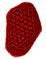
Protein Imager rendering Example. HIV Viral capsid
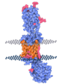
Protein Imager rendering Example. Membrane protein
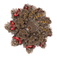
Protein Imager rendering Example. Protein complex


References
- ↑ Tomasello, Gianluca; Armenia, Ilaria; Molla, Gianluca (2020-05-01). "The Protein Imager: a full-featured online molecular viewer interface with server-side HQ-rendering capabilities" (in en). Bioinformatics 36 (9): 2909–2911. doi:10.1093/bioinformatics/btaa009. ISSN 1367-4803. PMID 31930403. https://academic.oup.com/bioinformatics/article/36/9/2909/5701652.
- ↑ "3D Protein Imager a PyMOL/Qutemol web alternative" (in en-US). 2024-02-06. https://bcrf.biochem.wisc.edu/2024/02/06/3d-protein-imager-a-pymol-qutemol-web-alternative/.
- ↑ "About Protein Imager" (in en-GB). https://3dproteinimaging.com/about-protein-imager/.
- ↑ RCSB, Protein Data Bank. "Molecular Graphics Software" (in en-US). https://www.rcsb.org/docs/additional-resources/molecular-graphics-software.
- ↑ "Illustrate: Non-photorealistic Biomolecular Illustration" (in en-US). https://ccsb.scripps.edu/illustrate/.
- ↑ "Protein Imager". https://my.labbrowser.com/store/app/3-d-protein-imaging.
- ↑ "Visualizing and Comparing Molecular Structures in Google Colab" (in en). https://colab.research.google.com/github/pb3lab/ibm3202/blob/master/tutorials/lab02_molviz.ipynb#scrollTo=osOd5k9E03KV.
- ↑ "«The Protein Imager»: un visualizzatore molecolare dal gruppo di ricerca «The Protein Factory 2.0» | Università degli studi dell'Insubria" (in it). https://archivio.uninsubria.it/notizie/%C2%AB-protein-imager%C2%BB-un-visualizzatore-molecolare-dal-gruppo-di-ricerca-%C2%AB-protein-factory-20%C2%BB.
- ↑ Noroozi, Fereshteh. "Exploring 3D Protein Imaging Web Service: A Step-by-Step Guide". https://www.youtube.com/watch?v=uqL3uSRmS38.
- ↑ Langendonk, R. Frèdi; Neill, Daniel R.; Fothergill, Joanne L. (2021). "The Building Blocks of Antimicrobial Resistance in Pseudomonas aeruginosa: Implications for Current Resistance-Breaking Therapies". Frontiers in Cellular and Infection Microbiology 11. doi:10.3389/fcimb.2021.665759. ISSN 2235-2988. PMID 33937104.
- ↑ Leitão, Ana Lúcia; Enguita, Francisco J. (2022-01-18). "A Structural View of miRNA Biogenesis and Function" (in en). Non-Coding RNA 8 (1): 10. doi:10.3390/ncrna8010010. ISSN 2311-553X. PMID 35202084.
- ↑ Ismail, Helene; Shakkour, Zaynab; Tabet, Maha; Abdelhady, Samar; Kobaisi, Abir; Abedi, Reem; Nasrallah, Leila; Pintus, Gianfranco et al. (2020-10-01). "Traumatic Brain Injury: Oxidative Stress and Novel Anti-Oxidants Such as Mitoquinone and Edaravone" (in en). Antioxidants 9 (10): 943. doi:10.3390/antiox9100943. ISSN 2076-3921. PMID 33019512.
- ↑ Ovejero, Jesus G.; Armenia, Ilaria; Serantes, David; Veintemillas-Verdaguer, Sabino; Zeballos, Nicoll; López-Gallego, Fernando; Grüttner, Cordula; de la Fuente, Jesús M. et al. (2021-09-08). "Selective Magnetic Nanoheating: Combining Iron Oxide Nanoparticles for Multi-Hot-Spot Induction and Sequential Regulation" (in en). Nano Letters 21 (17): 7213–7220. doi:10.1021/acs.nanolett.1c02178. ISSN 1530-6984. PMID 34410726. Bibcode: 2021NanoL..21.7213O.
 |  |

