Biology:Body-stalk
From HandWiki
| Body-stalk | |
|---|---|
 Diagram showing the expansion of amnion and delimitation of the umbilical cord | |
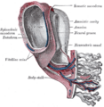
 Section through the embryo | |
| Details | |
| Identifiers | |
| Latin | Pedunculus truncalis |
| Anatomical terminology | |
The body-stalk, also known as the allantoic stalk,[1] is a band of mesoderm that connects the caudal end of the embryo to the chorion in development. With the formation of the caudal fold, the body-stalk assumes a ventral position; a diverticulum of the yolk-sac extends into the tail fold and is termed the hindgut. With continued development, the body-stalk is later replaced by the umbilical cord.
Body stalk anomalies occur in approximately 1 in 15,000 births.[2] They are due to defects in the formation of cephalic, caudal, and lateral embryonic body folds,[3] that result in a reduced or absent umbilical cord.[4]
Images
-
 Human embryo—length, 2 mm. Dorsal view, with the amnion laid open. X 30.
 -
 Human embryo of 2.6 mm.
 -
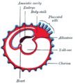 Diagram showing later stage of allantoic development with commencing constriction of the yolk-sac.
 -
 Model of human embryo 1.3 mm. long.
 -
 Section through ovum imbedded in the uterine decidua.
 -
 Embryo between eighteen and twenty-one days.
 -
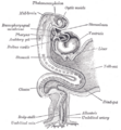 Human embryo about fifteen days old. Brain and heart represented from right side. Digestive tube and yolk sac in median section.


References
- ↑ Arthur Robinson (1913). Cunningham's Textbook of Anatomy. William Wood. pp. 54. https://books.google.com/books?id=my9AAAAAYAAJ&pg=PA54.
- ↑ Asim Kurjak (30 June 2013). Donald School Textbook of Transvaginal Sonography. JP Medical Ltd. pp. 28. ISBN 978-93-5090-473-2. https://books.google.com/books?id=xYuIWZE1Se4C&pg=PA28.
- ↑ Diana W. Bianchi; Timothy M. Crombleholme; Mary E. D'Alton (1 January 2000). Fetology: Diagnosis & Management of the Fetal Patient. McGraw Hill Professional. ISBN 978-0-8385-2570-8. https://books.google.com/books?id=53Csy-mr1bsC.
- ↑ Kocherla, K; Kumari, V; Kocherla, PR (January 2015). "Prenatal diagnosis of body stalk complex: A rare entity and review of literature.". The Indian Journal of Radiology & Imaging 25 (1): 67–70. doi:10.4103/0971-3026.150162. PMID 25709170.
External links
- Swiss embryology (from UL, UB, and UF) hdisqueembry/triderm0
 |
