Biology:Cerebrospinal fibers
From HandWiki
Short description: Derived from the cells of the motor area of the cerebral cortex
| Cerebrospinal fibers | |
|---|---|
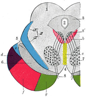
 Coronal section through mid-brain.
| |
| Details | |
| Identifiers | |
| Latin | Fibrae cerebrospinales |
| Anatomical terms of neuroanatomy | |
The cerebrospinal fibers, derived from the cells of the motor area of the cerebral cortex,[1] occupy the middle three-fifths of the base; they are continued partly to the nuclei of the motor cranial nerves, but mainly into the pyramids of the medulla oblongata.
References
- ↑ Chen, Hong; Zhang, Yan; Yang, Zhijun; Zhang, Hongtian (5 April 2013). "Human umbilical cord Wharton's jelly-derived oligodendrocyte precursor-like cells for axon and myelin sheath regeneration". Neural Regeneration Research 8 (10): 890–899. doi:10.3969/j.issn.1673-5374.2013.10.003. PMID 25206380.
 |

