Biology:Frontopontine fibers

From HandWiki
Frontopontine fibers
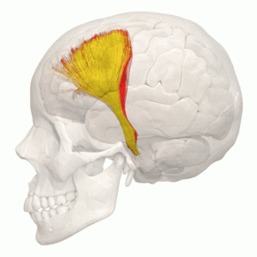
Tractography - Frontopontine tract - animation 1.gif
Tractography of frontopontine fibers
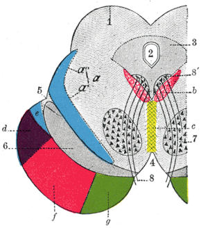
Gray710.png
Coronal section through mid-brain.
  1. Corpora quadrigemina
  2. Cerebral aqueduct
  3. Central gray stratum
  4. Interpeduncular space
  5. Sulcus lateralis
  6. Substantia nigra
  7. Red nucleus of tegmentum
  8. Oculomotor nerve, with 8’, its nucleus of origin
  1. Lemniscus (in blue) with a’ the medial lemniscus and a" the lateral lemniscus
  2. Medial longitudinal fasciculus
  3. Raphé
  4. Temporopontine fibers
  5. Portion of medial lemniscus, which runs to the lentiform nucleus and insula
  6. Cerebrospinal fibers
  7. Frontopontine fibers
Details
Identifiers
Latinfibrae frontopontinae
Anatomical terms of neuroanatomy

The frontopontine fibers[1] are situated in the medial fifth of the base of the cerebral peduncles; they arise from the cells of the frontal lobe and then pass through the anterior limb of internal capsule at last end in the nuclei of the pons.

The frontopontine tract (tractus frontopontinus) refers to the combination of the fibers.

See also

  • Paramedian pontine reticular formation

References

  1. Kamali A, Kramer LA, Frye RE, Butler IJ, Hasan KM. Diffusion tensor tractography of the human brain cortico-ponto-cerebellar pathways: a quantitative preliminary study. J Magn Reson Imaging. 2010 Oct;32(4):809-17. doi: 10.1002/jmri.22330. PMID 20882611

External links