Biology:Diagonal band of Broca
| Diagonal band of Broca | |
|---|---|
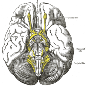
 Base of brain. (Diagonal band of Broca not labeled, but anterior perforated substance labeled at center.) | |
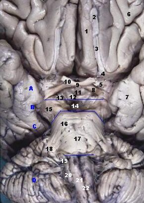
 Human brainstem, anterior view. (Diagonal band of Broca not labeled, but anterior perforated substance is #5.) | |
| Details | |
| Identifiers | |
| Latin | stria diagonalis |
| Anatomical terms of neuroanatomy | |
The diagonal band of Broca interconnects the amygdala and the septal area. It is one of the olfactory structures. It is situated upon the inferior aspect of the brain.[1] It forms the medial margin of the anterior perforated substance.[2]
It was described by the French neuroanatomist Paul Broca.[3]
Structure
It consists of fibers that are said to arise in the parolfactory area, the gyrus subcallosus and the anterior perforated substance, and course backward in the longitudinal striae to the dentate gyrus and the hippocampal region.[citation needed]
This is a cholinergic bundle of nerve fibers posterior to the anterior perforated substance. It interconnects the subcallosal gyrus in the septal area with the hippocampus and lateral olfactory area.[citation needed]
Nuclei
Two structures are often described in this brain regions, namely the nuclei of the vertical and horizontal limbs of the diagonal band of Broca (nvlDBB and nhlDBB, respectively). nvlDBB projects to the hippocampal formation through the fornix and it is the second largest assembly of cholinergic neurons in the basal forebrain whereas nhlDBB projects to the olfactory bulb and it does not have a significant population of cholinergic neurons.[3]
Development
It is one of the basal forebrain structures that are derived from the ventral telencephalon during development.[2]
Function
Along with the septum pellucidum and medial septal nucleus, the diagonal band of Broca is believed to be involved in the generation of theta waves in the hippocampus.[4] It also inhibits magnocellular neurosecretory cells via GABA interneurons.[5]
Its behavior can be altered by nerve growth factor.[6]
Pathology
A significant nvlDBB neuronal loss is seen in Lewy body dementia.[3]
References
- ↑ Gould, Douglas J.; Brueckner-Collins, Jennifer K.; Fix, James Douglas (2015). High-Yield Neuroanatomy (5th ed.). Philadelphia: Wolters Kluwer. pp. 4. ISBN 978-1-4511-9343-5.
- ↑ 2.0 2.1 Fix, James D. (2002). Neuroanatomy. Hagerstwon, MD: Lippincott Williams & Wilkins. pp. 338. ISBN 0-7817-2829-0. https://archive.org/details/neuroanatomy0003fixj.
- ↑ 3.0 3.1 3.2 Liu, A. K. L.; Lim, E. J.; Ahmed, I.; Chang, R. C.‐C.; Pearce, R. K. B.; Gentleman, S. M. (December 2018). "Review: Revisiting the human cholinergic nucleus of the diagonal band of Broca". Neuropathology and Applied Neurobiology 44 (7): 647–662. doi:10.1111/nan.12513. ISSN 0305-1846. PMID 30005126.
- ↑ O'Keefe, John; Andersen, Per; Morris, Richard; David Amaral; Tim Bliss (2007). The hippocampus book. Oxford [Oxfordshire]: Oxford University Press. pp. 480. ISBN 978-0-19-510027-3.
- ↑ Brown, Colin H. (2016). "Magnocellular Neurons and Posterior Pituitary Function" (in en). Comprehensive Physiology (American Cancer Society) 6 (4): 1701–1741. doi:10.1002/cphy.c150053. ISBN 9780470650714. PMID 27783857.
- ↑ "Chronic exposure to nerve growth factor increases acetylcholine and glutamate release from cholinergic neurons of the rat medial septum and diagonal band of Broca via mechanisms mediated by p75NTR". J. Neurosci. 28 (6): 1404–9. February 2008. doi:10.1523/JNEUROSCI.4851-07.2008. PMID 18256260.
 |

