Biology:Pacinian corpuscle
| Pacinian corpuscle | |
|---|---|
 Pacinian corpuscle, with its system of capsules and central cavity. a. Arterial twig, ending in capillaries, which form loops in some of the intercapsular spaces, and one penetrates to the central capsule. b. The fibrous tissue of the stalk. n. Nerve tube advancing to the central capsule, there losing its white matter and stretching along the axis to the opposite end, where it ends by a tuberculated enlargement. | |
 Pacinian corpuscle labeled at bottom | |
| Details | |
| Location | Skin |
| Identifiers | |
| Latin | corpusculum Pacinian |
| Anatomical terms of microanatomy | |
The Pacinian corpuscle (also lamellar corpuscle, or Vater–Pacini corpuscle)[1] is a low-threshold mechanoreceptor responsive to vibration or pressure, found in the skin and other internal organs.[2] In the skin it is one of the four main types of cutaneous receptors.
The corpuscles are present in skin notably on both surfaces of the hands and feet, arms, and neck.[3] Pacinian corpuscles are also found on bone periosteum, joint capsules, the pancreas and other internal organs, the breast, genitals,[4] and lymph nodes.[5]
Structure
Pacinian corpuscles are larger and fewer in number than Meissner's corpuscles, Merkel cells and Ruffini's corpuscles.[6] They may measure up to 2 mm in length, and nearly 1 mm in diameter.[7] They are oval, spherical, or irregularly coiled in shape. Larger ones are visible to the naked eye.[3] They have large receptive fields - as large as half of the palm.[7] In the skin, the corpuscles are situated deep within the dermis.[7]
Axon terminal
Each corpuscle is associated with a myelinated axon;[3] these are some of the largest and fastest-conducting sensory axons arising from the skin.[7]
Towards the center of the corpuscle, the axon loses its sheaths, ending as with a slight bulge at the center of the corpuscle. This axon terminal issues brief projections of unknown functional significance into gaps between the surrounding innermost lamellae; large mitochondria and small vessels aggregate near these projections.[3]
Capsule
The capsule consists of 20-70 concentrically-arranged connective tissue lamellae around the axon terminal at its center, forming a structure much like an onion.[7] The capsule consists of fibroblasts and fibrous connective tissue (mainly Type IV and Type II collagen network), separated by gelatinous material, more than 92% of which is water.[8] It presents a whorled pattern on micrographs. If the corpuscle's capsule is experimentally removed, the divested axon terminal becomes slowly adapting. The capsule is therefore responsible for the corpuscle's selectivity for high-frequency stimuli. This is a result of the slippery lamellae sliding past each other when the corpuscle is structurally deformed by external pressure so that effects of sustained pressure are soon dissipated by the lamellae, abolishing deformation of the central axon terminal itself.[7] The capsule thus acts as a physiological high-pass filter.[3]
Function
This section relies too much on references to primary sources. (September 2024) (Learn how and when to remove this template message) |
Pacinian corpuscles are rapidly adapting phasic receptors that detect gross pressure changes and vibrations in the skin.[9] Pacinian corpuscles have a large receptive field on the skin's surface with an especially sensitive center.[6]
The corpuscles are especially sensitive to vibrations, which they can sense even centimeters away.[6] Their optimal sensitivity is 250 Hz, and this is the frequency range generated upon fingertips by textures made of features smaller than 1 μm.[10][11] Pacinian corpuscles respond when the skin is rapidly indented but not when the pressure is steady (due to the capsule).[6] It is thought that they respond to high-velocity changes in joint position. They have also been implicated in detecting the location of touch sensations on handheld tools.[12]
Sensory transduction
Pacinian corpuscles sense stimuli due to the deformation of their lamellae in the capsule and inner core, which in turn press on the membrane (axolemma) of the sensory neuron and causes it to bend or stretch.[13] The external stimulus (deformation of or force on the external surface of the capsule) reaches axolemma of the terminal neurite through a complex mechanical filtration process. The internal lamellar spacing, number of lamella present in the capsule and the biomechanical properties of lamellae and the interlamellar fluid governs the characteristics of this mechanical filter acting on the external stimulus.[14][15][16] When the axolemma are deformed by the filtered stumulus, due to either application or release of the external stimulus, a generator or receptor potential is created as it physically deforms the plasma membrane of axon terminal, making it "leak" different cations through mechanosensitive channels which initiates the receptor potential. This initial receptor potential is potentiated by voltage-activated ion channels present in the inner-core of the corpuscle. Finally, the receptor potential is modulated to neural spikes or action potential with the help of opening of sodium ion channels present at the first Ranvier's Node of the axon.[17]
Due to generation of receptor potential in the receptive area of the neurite (especially near the heminode or half-node of the axon) the potential at the first Ranvier's node can reach certain threshold, triggering nerve impulses or action potentials at the first node of Ranvier. The first Ranvier's node of the myelinated section of the neurite is often found inside the capsule [18]. This impulse is then transferred along the axon from node to node with the use of sodium channels and sodium/potassium pumps in the axon membrane.
Surface vibrations
The Pacinian corpuscles in elephant feet have been suggested to enable seismic communication.[19] The Pacinian corpuscles in mice can detect taps on a branch 2.5 meters away.[20]
History
Pacinian corpuscles were the first cellular sensory receptor ever observed. They were first reported by German anatomist and botanist Abraham Vater and his student Johannes Gottlieb Lehmann in 1741, but ultimately named after Italian anatomist Filippo Pacini, who rediscovered them in 1835.[21][22] John Shekleton, a curator of the Royal College of Surgeons in Ireland, also discovered them before Pacini, but his results were published later.[21] Similar to Pacinian corpuscles, Herbst corpuscles and Grandry corpuscles are found in bird species.
Additional images
-
 Diagrammatic sectional view of the skin (magnified)
 -
 Schema (German)
 -
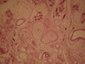 Light micrograph showing three corpuscles in the center of the field
 -
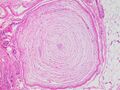 Micrograph of a Pacinian corpuscle


See also
- Pallesthesia
- List of human anatomical parts named after people
- Pacinian neuroma – a very rare benign tumor of Pacinian corpuscles
- Rayleigh wave
References
- ↑ Germann, C.; Sutter, R.; Nanz, D. (June 2021). "Novel observations of Pacinian corpuscle distribution in the hands and feet based on high-resolution 7-T MRI in healthy volunteers.". Skeletal Radiology 50 (6): 1249–1255. doi:10.1007/s00256-020-03667-7. PMID 33156397.
- ↑ Cobo, R; García-Piqueras, J; Cobo, J; Vega, JA (10 January 2021). "The Human Cutaneous Sensory Corpuscles: An Update.". Journal of Clinical Medicine 10 (2): 227. doi:10.3390/jcm10020227. PMID 33435193.
- ↑ 3.0 3.1 3.2 3.3 3.4 Standring, Susan (2020). Gray's Anatomy: The Anatomical Basis of Clinical Practice (42th ed.). New York: Elsevier. pp. 63–64. ISBN 978-0-7020-7707-4. OCLC 1201341621.
- ↑ Clark, Mary Ann; Douglas, Matthew; Choi, Jung (28 March 2018). "36.2 Somatosensation - Biology 2e | OpenStax" (in English). https://openstax.org/books/biology-2e/pages/36-2-somatosensation.
- ↑ Feito, J.; Cobo, J.L.; Santos-Briz, A.; Vega, J.A. (2017). "Pacinian Corpuscles in Human Lymph Nodes". The Anatomical Record (Wiley) 300 (12): 2233–2238. doi:10.1002/ar.23679. ISSN 1932-8486.
- ↑ 6.0 6.1 6.2 6.3 Principles of Neural Science. New York, NY: McGraw-Hill, Health Professions Division. 2000. ISBN 0-8385-7701-6. https://archive.org/details/isbn_9780838577011.
- ↑ 7.0 7.1 7.2 7.3 7.4 7.5 Bear, Mark F.; Connors, Barry W.; Paradiso, Michael A. (2016). Neuroscience: Exploring the Brain (4th ed.). Philadelphia: Wolters Kluwer. pp. 417–420. ISBN 978-0-7817-7817-6.
- ↑ Cherepnov, V.L.; Chadaeva, N.I. (1981). "Some characteristics of soluble proteins of Pacinian corpuscles". Bulletin of Experimental Biology and Medicine 91 (3): 346–348. doi:10.1007/BF00839370. PMID 7248510.
- ↑ Purves, Dale; Augustine, George J.; Fitzpatrick, David; Katz, Lawrence C.; LaMantia, Anthony-Samuel; McNamara, James O.; Williams, S. Mark (2001). "Cutaneous and Subcutaneous Somatic Sensory Receptors" (in en). Sinauer Associates. https://www.ncbi.nlm.nih.gov/books/NBK11162/.
- ↑ Scheibert, J; Leurent, S; Prevost, A; Debrégeas, G (2009). "The role of fingerprints in the coding of tactile information probed with a biomimetic sensor". Science 323 (5920): 1503–6. doi:10.1126/science.1166467. PMID 19179493. Bibcode: 2009Sci...323.1503S.
- ↑ Skedung, Lisa, Martin Arvidsson, Jun Young Chung, Christopher M. Stafford, Birgitta Berglund, and Mark W. Rutland. 2013. "Feeling Small: Exploring the Tactile Perception Limits." Sci. Rep. 3 (September 12). doi:10.1038/srep02617.
- ↑ Sima, Richard (23 December 2019). "The Brain Senses Touch beyond the Body". Scientific American. https://www.scientificamerican.com/article/the-brain-senses-touch-beyond-the-body/.
- ↑ Klein, Stephen B.; Michael Thorne, B. (2006-10-03). Biological Psychology. Macmillan. ISBN 9780716799221. https://books.google.com/books?id=p4fNxxuuOgwC&q=bend+pacinian+corpuscle&pg=PA244.
- ↑ Biswas, Abhijit; Manivannan, M.; Srinivasan, Mandyam A. (2015). "Multiscale Layered Biomechanical Model of the Pacinian Corpuscle". IEEE Transactions on Haptics 8 (1): 31–42. doi:10.1109/TOH.2014.2369416. PMID 25398182. https://zenodo.org/record/894776.
- ↑ Hubbard, S. J. (1958). "A study of rapid mechanical events in a mechanoreceptor". The Journal of Physiology 141 (2): 198-218. doi:10.1113/jphysiol.1958.sp005968. http://jp.physoc.org/content/141/2/198.short.
- ↑ Loewenstein, W. R.; Skalak, R. (1966). "Mechanical transmission in a Pacinian corpuscle. An analysis and a theory". The Journal of Physiology 182 (2): 346-378. doi:10.1113/jphysiol.1966.sp007827. http://jp.physoc.org/content/182/2/346.abstract.
- ↑ Biswas, Abhijit; Manivannan, M.; Srinivasan, Mandyam A. (2015). "Vibrotactile sensitivity threshold: Nonlinear stochastic mechanotransduction model of the Pacinian corpuscle". IEEE Transactions on Haptics 8 (1): 102–113. doi:10.1109/TOH.2014.2369422. PMID 25398183. https://zenodo.org/record/894772.
- ↑ Diamond, J.; Gray, J. A. B.; Sato, M. (1956). "The site of initiation of impulses in Pacinian corpuscles". The Journal of Physiology 133 (1): 54-67. doi:10.1113/jphysiol.1956.sp005566. PMID 13346634. https://pmc.ncbi.nlm.nih.gov/articles/PMC1359137/.
- ↑ Bouley, D. M.; Alarcón, C. N.; Hildebrandt, T.; O’Connell-Rodwell, C. E. (2007). "The distribution, density and three-dimensional histomorphology of Pacinian corpuscles in the foot of the Asian elephant (Elephas maximus) and their potential role in seismic communication". Journal of Anatomy (Wiley) 211 (4): 428–435. doi:10.1111/j.1469-7580.2007.00792.x. ISSN 0021-8782.
- ↑ Turecek, Josef; Ginty, David D. (2024). "Coding of self and environment by Pacinian neurons in freely moving animals". Neuron (Elsevier BV) 112 (19): 3267–3277.e6. doi:10.1016/j.neuron.2024.07.008. ISSN 0896-6273.
- ↑ 21.0 21.1 Bentivoglio, Marina; Pacini, Paolo (1995). "Filippo Pacini: a determined observer" (in en). Brain Research Bulletin 38 (2): 161–165. doi:10.1016/0361-9230(95)00083-Q. PMID 7583342. https://linkinghub.elsevier.com/retrieve/pii/036192309500083Q.
- ↑ Cauna, N.; Mannan, G. (1958). "The structure of human digital pacinian corpuscles (corpus cula lamellosa) and its functional significance". Journal of Anatomy 92 (1): 1–20. ISSN 0021-8782. PMID 13513492.
External links
- Virginia Commonwealth University
- Anatomy Atlases - Microscopic Anatomy, plate 06.124
- Medical Illustration
 |  |

