Biology:Perifovea
From HandWiki
Short description: Region in the retina

Perifovea is a region in the retina that circumscribes the parafovea and fovea and is a part of the macula lutea.[1] The perifovea is a belt that covers a 10° radius around the fovea and is 1.5 mm wide.[2][3] The perifovea ends when the Henle's fiber layer disappears and the ganglion cells are one-layered.[4]
Additional images
-
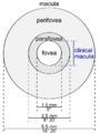 Schematic diagram of the macula lutea of the retina, showing perifovea, parafovea, fovea, and clinical macula
 -
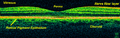 Time-Domain OCT of the macular area of a retina at 800 nm, axial resolution 3 µm
 -
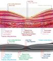
 Spectral-Domain OCT macula cross-section scan.
 -
 macula histology (OCT)
 -
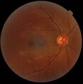 A fundus photograph showing the macula as a spot to the left. The optic disc is the area on the right where blood vessels converge. The grey, more diffuse spot in the centre is a shadow artifact.


See also
- Eye movements in reading
- Fixation (visual)
- Optical coherence tomography (OCT)
References
- ↑ Myron Yanoff; Jay S. Duker (6 November 2013). Ophthalmology: Expert Consult: Online and Print. Elsevier Health Sciences. pp. 421. ISBN 978-1-4557-5001-6. https://books.google.com/books?id=XfABAgAAQBAJ&pg=PA421.
- ↑ Jasjit S. Suri (2008). Image Modeling of the Human Eye. Artech House. pp. 133. ISBN 978-1-59693-209-8. https://books.google.com/books?id=ECXvpoWDotMC&pg=PA133.
- ↑ Vito Roberto (10 November 1993). Intelligent Perceptual Systems: New Directions in Computational Perception. Springer. pp. 347. ISBN 978-3-540-57379-1. https://books.google.com/books?id=9t1vHQxB9JIC&pg=PA347.
- ↑ Louis E. Probst; Julie H. Tsai; George Goodman (OD.) (2012). Ophthalmology: Clinical and Surgical Principles. SLACK Incorporated. pp. 28. ISBN 978-1-55642-735-0. https://books.google.com/books?id=QilKd2nvl8cC&pg=PA28.
| Fibrous tunic (outer) |
|  | |||||
|---|---|---|---|---|---|---|---|
| Uvea/vascular tunic (middle) |
| ||||||
| Retina (inner) |
| ||||||
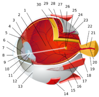
| Anatomical regions of the eye |
| ||||||
| Other | |||||||
 |  |
