Biology:Supraesophageal ganglion
| Look up supraesophageal in Wiktionary, the free dictionary. |
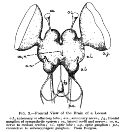
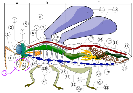
The supraesophageal ganglion (also "supraoesophageal ganglion", "arthropod brain" or "microbrain"[1]) is the first part of the arthropod, especially insect, central nervous system. It receives and processes information from the first, second, and third metameres. The supraesophageal ganglion lies dorsal to the esophagus and consists of three parts, each a pair of ganglia that may be more or less pronounced, reduced, or fused depending on the genus:
- The protocerebrum, associated with the eyes (compound eyes and ocelli).[2] Directly associated with the eyes is the optic lobe, as the visual center of the brain.
- The deutocerebrum processes sensory information from the antennae.[2][3] It consists of two parts, the antennal lobe and the dorsal lobe.[3][4][5] The dorsal lobe also contains motor neurons which control the antennal muscles.[6]
- The tritocerebrum integrates sensory inputs from the previous two pairs of ganglia.[2] The lobes of the tritocerebrum split to circumvent the esophagus and begin the subesophageal ganglion.
The subesophageal ganglion continues the nervous system and lies ventral to the esophagus. Finally, the segmental ganglia of the ventral nerve cord are found in each body segment as a fused ganglion; they provide the segments with some autonomous control.
A locust brain dissection to expose the central brain and carry out electro-physiology recordings can be seen here.[7]
See also
References
- ↑ Makoto Mizunami, Fumio Yokohari, Masakazu Takahata (1999). "Exploration into the Adaptive Design of the Arthropod "Microbrain"". Zoological Science 16 (5): 703–709. doi:10.2108/zsj.16.703.
- ↑ 2.0 2.1 2.2 Meyer, John R.. "The Nervous System". General Entomology course at North Carolina State University. Department of Entomology NC State University. https://projects.ncsu.edu/cals/course/ent425/library/tutorials/behavior/nervous.html.
- ↑ 3.0 3.1 Homberg, U; Christensen, T A; Hildebrand, J G (1989). "Structure and Function of the Deutocerebrum in Insects". Annual Review of Entomology 34: 477–501. doi:10.1146/annurev.en.34.010189.002401. PMID 2648971.
- ↑ "Invertebrate Brain Platform". RIKEN BSI Neuroinformatics Japan Center. https://invbrain.neuroinf.jp/modules/htmldocs/test/General/deutocerebrum.html.
- ↑ "Deutocerebrum". Flybrain. http://web.neurobio.arizona.edu/Flybrain/html/atlas/structures/deutocer.html.
- ↑ "Deutocerebrum". Invertebrate Brain Platform. https://invbrain.neuroinf.jp/modules/htmldocs/test/General/deutocerebrum.html. Chelicerata, with their missing antennae, have a very reduced (or absent) deutocerebrum.
- ↑ "Dissecting insect brain for in vivo electrophysiology". https://www.youtube.com/watch?v=Gav_rJhBfWY.
Further reading
- Erber, J.; Menzel, R. (1977). "Visual interneurons in the median protocerebrum of the bee". Journal of Comparative Physiology 121 (1): 65–77. doi:10.1007/bf00614181.
- Wong, Allan M., Jing W. Wang, and Richard Axel (2002). "Spatial Representation of the Glomerular Map in the Drosophila Protocerebrum". Cell 109 (2): 229–241. doi:10.1016/S0092-8674(02)00707-9. PMID 12007409.
- Malun, D.; Waldow, U.; Kraus, D.; Boeckh, J. (1993). "Connections between the deutocerebrum and the protocerebrum, and neuroanatomy of several classes of deutocerebral projection neurons in the brain of male Periplaneta americana". J. Comp. Neurol. 329 (2): 143–162. doi:10.1002/cne.903290202. PMID 8454728.
- Flanagan, Daniel; Mercer, Alison R. (1989). "Morphology and response characteristics of neurones in the deutocerebrum of the brain in the honeybeeApis mellifera". Journal of Comparative Physiology A 164 (4): 483–494. doi:10.1007/bf00610442.
- Childress, Steven A.; B. McIver, Susan (1984). "Morphology of the deutocerebrum of female Aedes aegypti (Diptera: Culicidae)". Canadian Journal of Zoology 62 (7): 1320–1328. doi:10.1139/z84-190.
- Technau, Gerhard (2008) (in nl). Brain development in Drosophila melanogaster. Advances in Experimental Medicine and Biology. 628. New York Austin, Tex: Springer Science+Business Media Landes Bioscience. doi:10.1007/978-0-387-78261-4. ISBN 978-0-387-78260-7. OCLC 314349837.
- Aubele, Elisabeth, and Nikolai Klemm (1977). "Origin, destination and mapping of tritocerebral neurons of locust". Cell and Tissue Research 178 (2): 199–219. doi:10.1007/bf00219048. PMID 66098.
- Chaudonneret, J. "Evolution of the insect brain with special reference to the so-called tritocerebrum." Arthropod brain. Wiley, New York (1987): 3-26.
- "Behavioral Neuroscience, lecture on Honey Bee and its behavior - Neuroanatomy". http://www.usdbiology.com/cliff/Courses/Behavioral%20Neuroscience/Bee/2%20Honey%20Bee%20Conditioned%20Drinking%20Neuroanatomy%20IV.html.
External links
- "Suboesophageal Ganglion". flybrain. http://web.neurobio.arizona.edu/Flybrain/html/atlas/structures/suoesg.html.
 |


