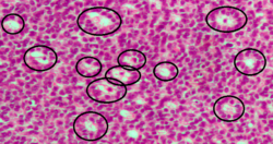
Medicine:Call-Exner bodies

Call–Exner bodies, giving a follicle-like appearance, are small eosinophilic fluid-filled punched out spaces between granulosa cells.[1] The granulosa cells are usually arranged haphazardly around the space.
They are pathognomonic for granulosa cell tumors.
Histologically, these tumors consists of monotonous islands of granulosa cells with "coffee-bean" nuclei. That same nuclear groove appearance noted in Brenner tumour, an epithelial-stromal ovarian tumor distinguishable by nests of transitional epithelial cells (urothelial) with longitudinal nuclear grooves (coffee bean nuclei) in abundant fibrous stroma. [1][2]
They are composed of membrane-packaged secretion of granulosa cells and have relations to the formation of liquor folliculi which are seen among closely arranged granulosa cells.
They are named for Emma Louise Call (1847-1937), an American physician, and Sigmund Exner.[3][4]
References
- ↑ thefreedictionary.com > Call–Exner body Citing: The American Heritage Medical Dictionary. Copyright 2007
- ↑ Ahr, A.; Arnold, G.; Göhring, U. J.; Costa, S.; Scharl, A.; Gauwerky, J. F. (July 1997). "Cytology of ascitic fluid in a patient with metastasizing malignant Brenner tumor of the ovary. A case report". Acta Cytologica 41 (4 Suppl): 1299–1304. doi:10.1159/000333524. ISSN 0001-5547. PMID 9990262.
- ↑ Archived version of the Call-Exner bodies page on Who Named It?
- ↑ Emma Louise Call and Sigmund Exner: Zur Kenntniss des Graafschen Follikels und des Corpus luteum beim Kaninchen. Sitzungsberichte der kaiserlichen Akademie der Wissenschaften. Mathematish naturwssenschaftliche Classe, Wien, 1875, 72: 321-328.
External links

