Medicine:Cavitation

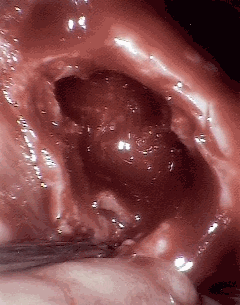
Cavitation is the formation of cavities, which are spaces or openings in the body. This process occurs in mammalian embryos and can also occur later on in fully developed organisms. During mammalian embryo development, cavitation is a routine process; however, the formation of cavities in fully developed organs, especially in lung tissue, is usually the sign of a severe medical condition or disease, such as tuberculosis. [1]
Developmental
Cavitation is a crucial process in the development of mammalian embryos. After fertilization, rapid cell division occurs which results in the formation of the morula, or a solid ball of cells. The morula is the precursor structure to the blastula, which is an animal embryo in the early stages of development. The morula consists of a cluster of internal cells covered by a layer of external cells. The internal cells become the inner cellular mass, which becomes the entire embryo.The external cells are destined to become a structure called the trophoblast, a layer of tissue on the inside of the embryo that provides it with nourishment. The trophoblast cells become extraembryonic structures necessary for development. After the initial formation of the morula, it does not have an interior space or cavity. Cavitation occurs to create a cavity on the inside of the morula. This process occurs when trophoblast cells, in other words the outside covering of the blastocyst, secretes fluid into the morula creating the blastocoel, the fluid filled cavity of the blastula. The formation of the blastocoel is a critical stage in the formation of the blastocyst, which is a blastula where some cellular differentiation has already occurred. [2]

Disease induced
When cavities form outside of development, it most commonly occurs in the lungs. This is usually the result of extensive damage done by a disease or medical condition. The disease itself does not directly cause the cavitation of the lung tissue. Instead, the disease induces necrosis the death of a group of cells or tissue, which in turn results in the formation of cavities within the infected area. Two diseases that are commonly associated with extensive necrosis and cavitation of lung tissue are Mycobacterium tuberculosis and Klebsiella pneumoniae. The formation of cavities due to tissue death creates an environment that allows the pathogen to expand in numbers and spread further.[3]
Detection and diagnosis
While cavity formation is a sign of severe tissue damage, these cavities can be used to aid in the diagnosis of the disease causing them. The most common imaging techniques used to document these cavities are chest x-ray and computed tomography (CT Scan). From these images, the wall thickness of the cavities is examined. Wall thickness helps point diagnosticians in the proper direction as to what the invading pathogen causing the cavitation might be. In addition to these scans, clinical and laboratory data is always taken and analyzed to confirm suspected diagnoses.[4]
References
- ↑ Mitchell, Richard Sheppard; Kumar, Vinay; Abbas, Abul K.; Fausto, Nelson. Robbins Basic Pathology. Philadelphia: Saunders. ISBN 1-4160-2973-7. 8th edition.
- ↑ Gilbert, SF (2000). Developmental Biology: Early Mammalian Development (6th ed.). Sunderland (MA).
- ↑ Gadkowski, L. Beth; Stout, Jason (21 April 2008). "Cavitary Pulmonary Disease". Clinical Microbiology Reviews.
- ↑ Gadkowski, L. Beth; Stout, Jason (21 April 2008). "Cavitary Pulmonary Disease". Clinical Microbiology Reviews.
