Medicine:Flexner–Wintersteiner rosette

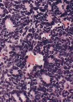
Flexner–Wintersteiner rosette is a spoke and wheel shaped cell formation seen in retinoblastoma and certain other ophthalmic tumors.[1] A rosette is a structure or formation resembling a rose, such as the clusters of polymorphonuclear leukocytes around a globule of lipid nuclear material, as observed in the test for disseminated lupus erythematosus.[1]
Characteristics
The tumor cells that form the Flexner–Wintersteiner rosette surround a central lumen containing small cytoplasmic extensions of the encircling cells. Unlike the center of the Homer Wright rosette, the central lumen is devoid of fiber-rich neuropil. Like the Homer Wright rosette, the Flexner–Wintersteiner rosette represents a specific form of tumor differentiation.[2][3][4][5] Electron microscopy reveals that the tumor cells forming the Flexner–Wintersteiner rosette have ultrastructural features of primitive photoreceptor cells.[6] Furthermore, the rosette lumen shows similar staining patterns as in rods and cones,[7] suggesting that Flexner–Wintersteiner rosettes represent a specific form of retinal differentiation. In addition to being a characteristic finding in retinoblastomas, Flexner–Wintersteiner rosettes may also be found in pinealoblastomas and medulloepitheliomas.[2]
History
Flexner–Wintersteiner rosettes were first described by Simon Flexner (1863–1946), a physician, scientist, administrator, and professor of experimental pathology at the University of Pennsylvania (1899–1903). Flexner noted characteristic clusters of cells in an infantile eye tumor which he called retinoepithelioma.[8][9][10] A few years later, in 1897, Austrian ophthalmologist Hugo Wintersteiner (1865–1946) confirmed Flexner’s observations and noted that the cell clusters resembled rods and cones.[11] These characteristic rosette formations were subsequently recognized as important features of retinoblastomas.
References
- ↑ 1.0 1.1 Definition of 'rosette', from The Free Dictionary. Retrieved 6 January 2010.
- ↑ 2.0 2.1 McLean IW, Burnier MN, Zimmerman LE, et al. Tumors of the retina. In: Atlas of tumor pathology: tumors of the eye and ocular adnexa. Washington, DC: Armed Forces Institute of Pathology; 1994:97–154
- ↑ Donoso LA, Shields CL, Lee EY-H. Immunohistochemistry of retinoblastoma. Ophthal Paediatr Genet 1989;10:3–32
- ↑ Vrabec T, Arbizo V, Adamus G, et al. Rod cell-specific antigens in retinoblastoma. Arch Ophthalmol 1989;107:1061–63
- ↑ Kivela T. Glycoconjugates in retinoblastoma: a lectin histochemical study of ten formalin-fixed and paraffin embedded tumours. Virchows Arch A 1987;410:471–79
- ↑ Ts’o MOM, Fine BS, Zimmerman LE. The Flexner–Wintersteiner rosettes in retinoblastoma. Arch Pathol 1969;88:664–71
- ↑ Zimmerman LE. Retinoblastoma and retinocytoma. In: Spencer WH, ed. Ophthalmic pathology: an atlas and textbook. Philadelphia: WB Saunders; 1985:1292–351
- ↑ Flexner S. A peculiar glioma (neuroepithelioma?) of the retina. Johns Hopkins Hosp Bull 1891;2:115–19
- ↑ Schatski SC. Simon Flexner. AJR Am J Roentgenol 1997;169:1395–96.
- ↑ Bendiner E. Simon Flexner: his "rock" was for the ages. Hosp Pract 1988;23:213–66
- ↑ Wintersteiner H. Die zellen der geschwulst. In: Das neuroepithelioma retinae: Eine anatomische und klinishe studie. Leipzig: Franz Deuticke; 1897:12–16

