Medicine:Parathyroid carcinoma
| Parathyroid carcinoma | |
|---|---|
 | |
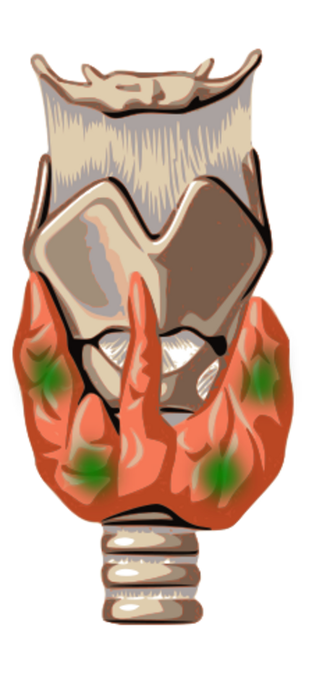
| Parathyroid gland anatomy(green marks) | |
| Specialty | Oncology, ENT surgery |
Parathyroid carcinoma is a rare cancer resulting in parathyroid adenoma to carcinoma progression.[1] It forms in tissues of one or more of the parathyroid glands (four pea-sized glands in the neck that make parathyroid hormone (PTH). PTH helps the body maintain normal levels of serum calcium by promoting calcium reabsorption from bone. It is antagonized by the hormone calcitonin, which prompts calcium storage.).
It is rare, with documented cases of less than one thousand since its first discovery in 1904;[2][3][4][5][6][7] and much less common than parathyroid adenoma. It can be difficult to excise.[8] The rate of occurrence of parathyroid carcinoma is between 0.5% to 5%[9][10][6][11]
Signs and symptoms
Most patients experience moderate to severe hypercalcemia and high parathyroid hormone levels. A large mass in the neck is often seen, and kidney and bone abnormalities are common.[1]
Risk factors
Parathyroid cancer occurs in midlife at the same rate in men and women. [12]
Conditions that appear to result in an increased risk of parathyroid cancer include multiple endocrine neoplasia type 1,[13] autosomal dominant familial isolated hyperparathyroidism[13] and hyperparathyroidism-jaw tumor syndrome[1] (which also is hereditary).[1] Parathyroid cancer has also been associated with external radiation exposure, but most reports describe an association between radiation and the more common parathyroid adenoma.[13]
Diagnosis
On Sestamibi parathyroid scan, intense radioactivity greater than submandibular gland on delayed image, no washout between early and delayed images, and high concentration of parathyroid hormone concentration in blood in those who age more than 40 years is suggestive of parathyroid carcinoma.[14] Some authors suggest high levels of HCG as a marker for parathyroid carcinoma in the right context.[15] However, other thyroid diseases such as multinodular goitre, Hashimoto thyroiditis, thyroid adenoma, and thyroid carcinoma also retains the radiotracer because of high metabolic nature of these diseases.[16] Thus, the final diagnosis always requires pathological examination of the tissue in question.
Treatment
Parathyroid carcinoma is sometimes diagnosed during surgery for primary hyperparathyroidism. If the surgeon suspects carcinoma based on severity or invasion of surrounding tissues by a firm parathyroid tumor, aggressive excision is performed, including the thyroid and surrounding tissues as necessary.[1]
Agents such as calcimimetics (for example, cinacalcet) are used to mimic calcium and are able to activate the parathyroid calcium-sensing receptor (making the parathyroid gland "think" we have more calcium than we actually do), therefore lowering the calcium level, in an attempt to decrease the hypercalcemia.[citation needed]
References
- ↑ 1.0 1.1 1.2 1.3 1.4 Hu MI, Vassilopoulou-Sellin R, Lustig R, Lamont JP. "Thyroid and Parathyroid Cancers" in Pazdur R, Wagman LD, Camphausen KA, Hoskins WJ (Eds) Cancer Management: A Multidisciplinary Approach. 11 ed. 2008.
- ↑ "Parathyroid carcinoma" (in English). Clinical Oncology 22 (6): 498–507. August 2010. doi:10.1016/j.clon.2010.04.007. PMID 20510594.
- ↑ "Parathyroid carcinoma: update and guidelines for management". Current Treatment Options in Oncology 13 (1): 11–23. March 2012. doi:10.1007/s11864-011-0171-3. PMID 22327883.
- ↑ "Parathyroid carcinoma". Otolaryngologic Clinics of North America. Thyroid and Parathyroid Surgery 43 (2): 441–53, xi. April 2010. doi:10.1016/j.otc.2010.01.011. PMID 20510726.
- ↑ "Parathyroid carcinoma". Otolaryngologic Clinics of North America. Parathyroids 37 (4): 845–54, x. August 2004. doi:10.1016/j.otc.2004.02.014. PMID 15262520.
- ↑ 6.0 6.1 "Parathyroid Cancer Cases: A Single Center's Experience" (in English). AACE Clinical Case Reports 2 (3): e221–e227. 2016-06-01. doi:10.4158/EP15857.CR. ISSN 2376-0605. https://www.aaceclinicalcasereports.com/article/S2376-0605(20)30590-3/abstract.
- ↑ "Predicting the presence of parathyroid carcinoma". Annals of Surgical Oncology 12 (7): 513–514. July 2005. doi:10.1245/ASO.2005.03.904. PMID 15952075.
- ↑ "Endocrine Pathology". http://library.med.utah.edu/WebPath/ENDOHTML/ENDO112.html.
- ↑ "Parathyroid carcinoma: clinical and pathologic features in 43 patients". Medicine 71 (4): 197–205. July 1992. doi:10.1097/00005792-199207000-00002. PMID 1518393. https://pubmed.ncbi.nlm.nih.gov/1518393.
- ↑ "Parathyroid carcinoma: challenges in diagnosis and treatment". Hematology/Oncology Clinics of North America. Rare Cancers 26 (6): 1221–1238. December 2012. doi:10.1016/j.hoc.2012.08.009. PMID 23116578.
- ↑ "Parathyroid carcinoma" (in English). Clinical Oncology 22 (6): 498–507. August 2010. doi:10.1016/j.clon.2010.04.007. PMID 20510594.
- ↑ "Parathyroid carcinoma" (in English). Clinical Oncology 22 (6): 498–507. August 2010. doi:10.1016/j.clon.2010.04.007. PMID 20510594.
- ↑ 13.0 13.1 13.2 "Parathyroid Cancer Treatment". National Cancer Institute. 11 March 2009. http://www.cancer.gov/cancertopics/pdq/treatment/parathyroid/HealthProfessional/page1.
- ↑ "Differential findings of tc-99m sestamibi dual-phase parathyroid scintigraphy between benign and malignant parathyroid lesions in patients with primary hyperparathyroidism". Nuclear Medicine and Molecular Imaging 45 (4): 276–284. December 2011. doi:10.1007/s13139-011-0103-y. PMID 24900018.
- ↑ "Human Chorionic Gonadotrophin (hCG) as a diagnostic test to differentiate between Parathyroid Carcinoma, Primary Benign Hyperparathyroidism and Secondary Hyperparathyroidism." (in en). Endocrine Abstracts (Bioscientifica) 56. 2018-05-08. doi:10.1530/endoabs.56.P214. https://www.endocrine-abstracts.org/ea/0056/ea0056p214.
- ↑ "Parathyroid imaging with Tc-99m sestamibi planar and SPECT scintigraphy". Radiographics 19 (3): 601–614. May 1999. doi:10.1148/radiographics.19.3.g99ma10601. PMID 10336191.
External links
- Parathyroid cancer entry in the public domain NCI Dictionary of Cancer Terms
- Parathyroid Carcinoma (Medscape eMedicine)
- Cancer Management Handbook: Thyroid and Parathyroid Cancers
| Classification | |
|---|---|
| External resources |
![]() This article incorporates public domain material from the U.S. National Cancer Institute document "Dictionary of Cancer Terms".
This article incorporates public domain material from the U.S. National Cancer Institute document "Dictionary of Cancer Terms".
 |

