Medicine:Submental triangle
From HandWiki
| Submental triangle | |
|---|---|
 Submental triangle | |
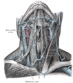
 Side of neck, showing chief surface markings (nerves are yellow, arteries are red) | |
| Details | |
| Identifiers | |
| Latin | Trigonum submentale |
| Anatomical terminology | |
The submental triangle (or suprahyoid triangle) is a division of the anterior triangle of the neck.
Boundaries
It is limited to:
- Lateral (away from the midline), formed by the anterior belly of the digastricus
- Medial (towards the midline), formed by the midline of the neck between the mandible and the hyoid bone
- Inferior (below), formed by the body of the hyoid bone
- Floor is formed by the mylohyoideus
- Roof is formed by Investing layer of deep cervical fascia
Contents
It contains one or two lymph glands, the submental lymph nodes (three or four in number) and Submental veins and commencement of anterior jugular veins.
(The contents of the triangle actually lie in the superficial fascia over the roof of submental triangle)
Additional images
-
 Muscles of the neck. Anterior view.
 -
 The veins of the neck, viewed from in front.
 -
 Front view of neck.
 -
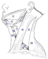 The triangles of the neck. (Anterior triangles to the left; posterior triangles to the right. Suprahyoid labeled at left.)


See also
- Anterior triangle of the neck
- Submental space
References
This article incorporates text in the public domain from the 20th edition of Gray's Anatomy (1918), Page 88 of Textbook of Anatomy; head, neck and brain by Vishram Singh
External links
- lesson5 at The Anatomy Lesson by Wesley Norman (Georgetown University) (necktriangle)
- lesson6 at The Anatomy Lesson by Wesley Norman (Georgetown University)
- Anatomy figure: 25:01-03 at Human Anatomy Online, SUNY Downstate Medical Center - "Identification of the subdivsions of the anterior triangle and corresponding borders."
- Anatomy photo:25:19-0101 at the SUNY Downstate Medical Center - "Anterior Triangle of the Neck: The Submental Triangle"
 |  |
