Biology:B2-adapt-app C
The C-terminal domain of Beta2-adaptin is a protein domain is involved in cell trafficking by aiding import and export of substances in and out of the cell.
Function
This is an adaptor protein which helps the formation of a clathrin coat around a vesicle.
Structure
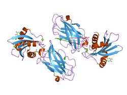
This entry represents a subdomain of the appendage (ear) domain of beta-adaptin from AP clathrin adaptor complexes. This domain has a three-layer arrangement, alpha-beta-alpha, with a bifurcated antiparallel beta-sheet.[1] This domain is required for binding to clathrin, and its subsequent polymerisation. Furthermore, a hydrophobic patch present in the domain also binds to a subset of D-phi-F/W motif-containing proteins that are bound by the alpha-adaptin appendage domain (epsin, AP180, eps15).[2]
Cell trafficking
Proteins synthesized on the ribosome and processed in the endoplasmic reticulum are transported from the Golgi apparatus to the trans-Golgi network (TGN), and from there via small carrier vesicles to their final destination compartment. These vesicles have specific coat proteins (such as clathrin or coatomer) that are important for cargo selection and direction of transport.[3] Clathrin coats contain both clathrin (acts as a scaffold) and adaptor complexes that link clathrin to receptors in coated vesicles. Clathrin-associated protein complexes are believed to interact with the cytoplasmic tails of membrane proteins, leading to their selection and concentration. The two major types of clathrin adaptor complexes are the heterotetrameric adaptor protein (AP) complexes, and the monomeric GGA (Golgi-localising, Gamma-adaptin ear domain homology, ARF-binding proteins) adaptors.[4][5]
AP (adaptor protein) complexes are found in coated vesicles and clathrin-coated pits. AP complexes connect cargo proteins and lipids to clathrin at vesicle budding sites, as well as binding accessory proteins that regulate coat assembly and disassembly (such as AP180, epsins and auxilin). There are different AP complexes in mammals. AP1 is responsible for the transport of lysosomal hydrolases between the TGN and endosomes.[6] AP2 associates with the plasma membrane and is responsible for endocytosis.[7] AP3 is responsible for protein trafficking to lysosomes and other related organelles.[8] AP4 is less well characterised. AP complexes are heterotetramers composed of two large subunits (adaptins), a medium subunit (mu) and a small subunit (sigma). For example, in AP1 these subunits are gamma-1-adaptin, beta-1-adaptin, mu-1 and sigma-1, while in AP2 they are alpha-adaptin, beta-2-adaptin, mu-2 and sigma-2. Each subunit has a specific function. Adaptins recognise and bind to clathrin through their hinge region (clathrin box), and recruit accessory proteins that modulate AP function through their C-terminal ear (appendage) domains. Mu recognises tyrosine-based sorting signals within the cytoplasmic domains of transmembrane cargo proteins.[9] One function of clathrin and AP2 complex-mediated endocytosis is to regulate the number of GABA(A) receptors available at the cell surface .[10]
More information about these proteins can be found at Protein of the Month: Clathrin .
References
- ↑ "Crystal structure of the alpha appendage of AP-2 reveals a recruitment platform for clathrin-coat assembly". Proc. Natl. Acad. Sci. U.S.A. 96 (16): 8907–12. August 1999. doi:10.1073/pnas.96.16.8907. PMID 10430869.
- ↑ "The structure and function of the beta 2-adaptin appendage domain". EMBO J. 19 (16): 4216–27. August 2000. doi:10.1093/emboj/19.16.4216. PMID 10944104.
- ↑ "COP and clathrin-coated vesicle budding: different pathways, common approaches". Curr. Opin. Cell Biol. 16 (4): 379–91. August 2004. doi:10.1016/j.ceb.2004.06.009. PMID 15261670.
- ↑ "Do different endocytic pathways make different synaptic vesicles?". Curr. Opin. Neurobiol. 17 (3): 374–80. June 2007. doi:10.1016/j.conb.2007.04.002. PMID 17449236.
- ↑ "Adaptins: the final recount". Mol. Biol. Cell 12 (10): 2907–20. October 2001. doi:10.1091/mbc.12.10.2907. PMID 11598180.
- ↑ "Adaptor protein complex 1 mediates the transport of lysosomal proteins from a Golgi-like organelle to peripheral vacuoles in the primitive eukaryote Giardia lamblia". Mol. Biol. Cell 15 (7): 3053–60. July 2004. doi:10.1091/mbc.E03-10-0744. PMID 15107467.
- ↑ "Differential requirements for AP-2 in clathrin-mediated endocytosis". J. Cell Biol. 162 (5): 773–9. September 2003. doi:10.1083/jcb.200304069. PMID 12952931.
- ↑ "Re-routing of the invariant chain to the direct sorting pathway by introduction of an AP3-binding motif from LIMP II". Eur. J. Cell Biol. 85 (6): 457–67. June 2006. doi:10.1016/j.ejcb.2006.02.001. PMID 16542748.
- ↑ "Dual interaction of synaptotagmin with mu2- and alpha-adaptin facilitates clathrin-coated pit nucleation". EMBO J. 19 (22): 6011–9. November 2000. doi:10.1093/emboj/19.22.6011. PMID 11080148.
- ↑ "Phospholipase C-related inactive protein is implicated in the constitutive internalization of GABAA receptors mediated by clathrin and AP2 adaptor complex". J. Neurochem. 101 (4): 898–905. May 2007. doi:10.1111/j.1471-4159.2006.04399.x. PMID 17254016.


