Biology:Cuneate nucleus
| Cuneate nucleus | |
|---|---|
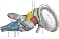
 Dissection of brain-stem. Dorsal view. (Label for "nucleus cuneatus" is on left, third from the bottom.) | |
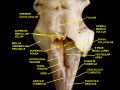
 Section of the medulla oblongata at about the middle of the olive. | |
| Details | |
| Identifiers | |
| Latin | nucleus cuneatus |
| Anatomical terms of neuroanatomy | |
One of the dorsal column nuclei, the cuneate nucleus is a wedge-shaped nucleus in the closed part of the medulla oblongata, in the brainstem. It contains cells that give rise to the cuneate tubercle, visible on the posterior aspect of the medulla. It lies laterally to the gracile nucleus and medial to the spinal trigeminal nucleus in the medulla.
Function
The cuneate nucleus is part of posterior column–medial lemniscus pathway, carrying fine touch and proprioceptive information from the upper body (above T6, except the face and ear—the information from the face and ear is carried by the primary sensory trigeminal nucleus) to the contralateral thalamus via the medial lemniscus.[citation needed]
Inputs
It receives direct input from the mechanoreceptors of the upper body as well as indirect input from them via the spinal cord. It is also subject to descending control from the central nervous system.[citation needed]
Pathology
It may be affected by vitamin E deficiency exhibiting neuroaxonal swelling.[1]
See also
- Fasciculus cuneatus
Additional images
-
 Decussation of pyramids.
 -
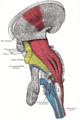
 Dissection of brain-stem. Lateral view.
 -
 Deep dissection of brain-stem. Lateral view.
 -
 Superior terminations of the posterior fasciculi of the medulla spinalis.
 -
 Diagram showing the course of the arcuate fibers.
 -
 The formatio reticularis of the medulla oblongata, shown by a transverse section passing through the middle of the olive.
 -
 Scheme showing the course of the fibers of the lemniscus; medial lemniscus in blue, lateral in red.
 -
 Transverse section passing through the sensory decussation.
 -
 Deep dissection of cortex and brain-stem.
 -
 The sensory tract.
 -
 Fourth ventricle.Posterior view.Deep dissection.


References
- ↑ Schmidt, Robert E.; Coleman, Bill D.; Nelson, James S. (1991). "Differential effect of chronic vitamin e deficiency on the development of neuroaxonal dystrophy in rat gracile/cuneate nuclei and prevertebral sympathetic ganglia". Neuroscience Letters 123 (1): 102–6. doi:10.1016/0304-3940(91)90168-S. PMID 2062445.
External links
- Stained brain slice images which include the "Cuneate nucleus" at the BrainMaps project
- NIF Search - Cuneate Nucleus via the Neuroscience Information Framework
 |
