Biology:Sucioplaca
| Sucioplaca | |
|---|---|

| |
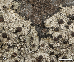
| Sucioplaca diplacia; scale bar = 2 mm | |
| Scientific classification | |
| Domain: | Eukaryota |
| Kingdom: | Fungi |
| Division: | Ascomycota |
| Class: | Lecanoromycetes |
| Order: | Teloschistales |
| Family: | Teloschistaceae |
| Genus: | Sucioplaca Bungartz, Søchting & Arup (2020) |
| Species: | S. diplacia
|
| Binomial name | |
| Sucioplaca diplacia (Ach.) Bungartz, Søchting & Arup (2020)
| |
| Synonyms[1] | |
| |
Sucioplaca is a single-species fungal genus in the family Teloschistaceae.[2][3] It contains Sucioplaca diplacia, a saxicolous (rock-dwelling) crustose lichen. It is common and widely distributed in the Caribbean, Central America, and the Galápagos Islands, where it grows on coastal rocks.[4]
Taxonomy
The genus Sucioplaca was formally proposed in 2020 by the lichenologists Frank Bungartz, Ulrik Søchting, and Ulf Arup. The name of the genus is derived from merging -placa, alluding to Caloplaca (the genus from which its species was reclassified), with sucio-, the Spanish term for "dirty". This reference is a nod to the dust-laden habitats near the ground where the sole known species of this genus is commonly found. Sucioplaca is in the subfamily Caloplacoideae of the family Teloschistaceae, where it is uniquely situated in a distinct clade, marking its isolated position within this grouping.[4] Sucioplaca diplacia was originally described by Erik Acharius in 1814 as a species of Lecanora.[5] In its taxonomic history, it has been proposed for inclusion in the genera Patellaria, Placodium, Caloplaca, and Callopisma.[1]
Description
The lichen Sucioplaca diplacia has a thallus, or main body, that varies from cracked ([[Glossary of lichen terms#{{biology:{1}}}|{{Biology:{1}}}]]) to a cracked and patchy (rimose-[[Glossary of lichen terms#{{biology:{1}}}|{{Biology:{1}}}]]) appearance. It ranges from thin to thick, generally bordered by a compact, blackish outline known as a prothallus. Although individual thalli can merge and blend with each other, they are usually distinct. The surface of Sucioplaca diplacia is dark to pale bluish-grey, occasionally with a pink tint, and has a smooth texture. It lacks a powdery coating ([[Glossary of lichen terms#{{biology:{1}}}|{{Biology:{1}}}]]) and features pimple-like (pustulate) soralia, which are structures for asexual reproduction, measuring 0.2–0.7 mm in diameter. These soralia contain pale green soredia, small granulular propagules, which may appear bluish-black when not eroded.[4]
Apothecia (fruiting bodies) are typically absent in Sucioplaca diplacia. If present, they are scattered or loosely grouped, sometimes deformed as if forming galls. They are seated ([[Glossary of lichen terms#{{biology:{1}}}|{{Biology:{1}}}]]) and small, usually less than 1 mm in diameter, with a [[Glossary of lichen terms#{{biology:{1}}}|{{Biology:{1}}}]] appearance (having a distinct margin), but can resemble other types. Initially pale, they darken with age, turning reddish-black. The margin of the apothecia is waxy, smooth, and initially pale, turning black over time. The [[Glossary of lichen terms#{{biology:{1}}}|{{Biology:{1}}}]], or central part, follows a similar colour transformation, from pale to deeply blackened, and is also smooth and epruinose. The [[Glossary of lichen terms#{{biology:{1}}}|{{Biology:{1}}}]], or upper layer above the spore-bearing tissue, is reddish-orange to violaceous and reacts yellowish-brown with the C chemical spot test. The hymenium, or spore-bearing layer, is clear (hyaline) and uncluttered. The structure surrounding the hymenium is poorly defined, with few hyaline hyphae (filamentous fungal cells).[4]
The [[Glossary of lichen terms#{{biology:{1}}}|{{Biology:{1}}}]] (the thallus-like layer around the hymenium) consists of three parts: a central part with abundant algae ([[Glossary of lichen terms#{{biology:{1}}}|{{Biology:{1}}}]]), a transitional part with small crystals dissolving in potassium hydroxide (K), and an outer part that becomes pigmented with age. The [[Glossary of lichen terms#{{biology:{1}}}|{{Biology:{1}}}]] and [[Glossary of lichen terms#{{biology:{1}}}|{{Biology:{1}}}]], supporting layers beneath the hymenium, are greyish and hyaline respectively. The asci, or spore sacs, are club-shaped ([[Glossary of lichen terms#{{biology:{1}}}|{{Biology:{1}}}]]) and of the Teloschistes type. Each ascus contains eight two-celled, broadly ellipsoid to almost spherical spores, with a thick septum. Pycnidia (asexual reproductive structures) have not been observed in this species.[4]
Secondary metabolites (lichen products) that occur in the thallus of Sucioplaca diplacia include atranorin, isofulgidin, vicanicin, and caloploicin. The expected results of standard chemical spot tests on the thallus are P+ (yellow), K+ (yellow), C–, and KC–; all spot tests on the apothecia are negative.[4]
References
- ↑ 1.0 1.1 "GSD Species Synonymy. Current Name: Sucioplaca diplacia (Ach.) Bungartz, Søchting & Arup, in Bungartz, Søchting & Arup, Plant and Fungal Systematics 65(2): 548 (2020)". Species Fungorum. https://www.speciesfungorum.org/GSD/GSDspecies.asp?RecordID=836962.
- ↑ "Sucioplaca". Species 2000: Naturalis, Leiden, the Netherlands. https://www.catalogueoflife.org/data/taxon/B2QGG.
- ↑ Wijayawardene, N.N.; Hyde, K.D.; Dai, D.Q.; Sánchez-García, M.; Goto, B.T.; Saxena, R.K. et al. (2022). "Outline of Fungi and fungus-like taxa – 2021". Mycosphere 13 (1): 53–453 [157]. doi:10.5943/mycosphere/13/1/2. https://www.researchgate.net/publication/358798332.
- ↑ 4.0 4.1 4.2 4.3 4.4 4.5 Bungartz, Frank; Søchting, Ulrik; Arup, Ulf (2020). "Teloschistaceae (lichenized Ascomycota) from the Galapagos Islands: a phylogenetic revision based on morphological, anatomical, chemical, and molecular data". Plant and Fungal Systematics 65 (2): 515–576. doi:10.35535/pfsyst-2020-0030.
- ↑ Acharius, E. (1814) (in la). Synopsis Methodica Lichenum. Lundin: Svanborg and Company. p. 217.
Wikidata ☰ entry
 |

