Biology:Trichosphaerium
| Trichosphaerium | |
|---|---|

| |
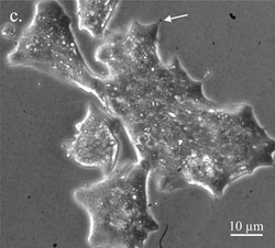
| Trichosphaerium sp. with dactylopodium (arrow) | |

| |
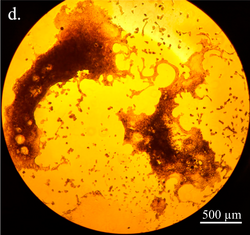
| Trichosphaerium in its giant amoeba form | |
| Scientific classification | |
| Domain: | Eukaryota |
| Phylum: | Amoebozoa |
| Class: | Tubulinea |
| Clade: | Corycidia |
| Order: | Trichosida Möbius, 1889 |
| Family: | Trichosphaeriidae Sheehan & Banner, 1973 |
| Genus: | Trichosphaerium Schneider, 1878 |
| Synonyms | |
| |
Trichosphaerium is a genus of amoebozoan protists that present extraordinary morphological transformations, both in size and shape, during their life cycle. They can present a test that may or may not be covered in spicules. They are related to the family Microcoryciidae, which contains other amoebae with tests, within the clade Corycidia of the phylum Amoebozoa.
Morphology
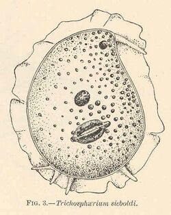
Trichosphaerium is a genus of amoebae characterized from other Amoebozoa by a multiporous test and a specialized non-motile pseudopodium, known as a dactylopodium, shaped like a digit. The dactylopodium is considered a sensory structure. Its morphology, behavior and life cycle are extraordinary in comparison with other protists. During its poorly understood life cycle, Trichosphaerium undergoes dramatic changes in shape and size. They can grow from as small as 10 µm to giant cell sizes of over 1 mm, observable by the naked eye. They can display such varied recognizable morphotypes that they can be easily mistaken with other species of amoebae.[2]

Controversial reports describe an alternation of two trophozoite stages within its life cycle: the "schizont", an amoeba surrounded by a test covered in flexible spicules, and the "gamont", an amoeba surrounded by a more flexible and fibrous test without spicules. According to studies written by Germany protozoologist Fritz Schaudinn in 1899, the gamont stage produces flagellated gametes, which fuse into a zygote to generate the schizont stage. Although both morphotypes have been observed and kept in laboratory cultures over the decades, this alternation of generations has never been observed in them, which adds a layer of complexity to the unusual, poorly understood behavior of these amoebae.[2]
Systematics

Trichosphaerium is the sole accepted genus of the family Trichosphaeriidae (sometimes written as Trichosidae)[3] and the order Trichosida.[1][4] The phylogenetic placement of Trichosphaerium has been controversial,[2] but most recent studies place it within the class Tubulinea of the phylum Amoebozoa.[1][5] In particular, since 2017, phylogenomic analyses of Amoebozoa recover a clade known as Corycidia, at the base of Tubulinea, containing both Trichosphaerium and amoebae of the family Microcoryciidae together.[4][6]
Synonyms
In 2016, American protozoologist Thomas Cavalier-Smith described the genus Atrichosa to comprise an undescribed species of Trichosphaerium, after considering that the type strain of this species does not belong to the genus Trichosphaerium but to a distinct, yet related, organism.[1] This change, however, was not accepted by the 2019 revision of eukaryotic classification, where Atrichosa is considered a junior synonym of Trichosphaerium "until the opposite is shown".[7] Another genus, Pontifex, is considered to be a synonym of Trichosphaerium, although with uncertainty.[1]
Species
Up to four species have been described within the genus, mainly based on the morphology of the spicules that cover their test.[2]
- Atrichosa algivora Cavalier-Smith, 2016 — described from the only strain of Trichosphaerium that has been genetically sequenced, ATCC 40318.[1] It is not accepted as a separate genus Atrichosa by other authors, but has not been formally merged back into Trichosphaerium.[7]
- Trichosphaerium micrum R.W. Angell, 1975[8]
- Trichosphaerium platyxyrum R.W. Angell, 1976[9]
- Trichosphaerium sieboldi Schneider, 1878
References
- ↑ 1.0 1.1 1.2 1.3 1.4 1.5 "187-gene phylogeny of protozoan phylum Amoebozoa reveals a new class (Cutosea) of deep-branching, ultrastructurally unique, enveloped marine Lobosa and clarifies amoeba evolution". Molecular Phylogenetics and Evolution 99: 275–296. June 2016. doi:10.1016/j.ympev.2016.03.023. PMID 27001604.
- ↑ 2.0 2.1 2.2 2.3 Tekle, Yonas I.; Tran, Hanh; Wang, Fang; Singla, Mandakini; Udu, Isimeme (2023). "Omics of an Enigmatic Marine Amoeba Uncovers Unprecedented Gene Trafficking from Giant Viruses and Provides Insights into Its Complex Life Cycle". Microbiology Research 14 (2): 656–672. doi:10.3390/microbiolres14020047. PMID 37752971.
- ↑ "The new higher level classification of eukaryotes with emphasis on the taxonomy of protists". The Journal of Eukaryotic Microbiology 52 (5): 399–451. 2005. doi:10.1111/j.1550-7408.2005.00053.x. PMID 16248873. http://doc.rero.ch/record/14409/files/PAL_E1847.pdf.
- ↑ 4.0 4.1 Kang, Seungho; Tice, Alexander K; Spiegel, Frederick W; Silberman, Jeffrey D; Pánek, Tomáš; Čepička, Ivan; Kostka, Martin; Kosakyan, Anush et al. (September 2017). "Between a Pod and a Hard Test: The Deep Evolution of Amoebae". Molecular Biology and Evolution 34 (9): 2258–2270. doi:10.1093/molbev/msx162. PMID 28505375.
- ↑ "Longamoebia is not monophyletic: Phylogenomic and cytoskeleton analyses provide novel and well-resolved relationships of amoebozoan subclades". Molecular Phylogenetics and Evolution 114: 249–260. September 2017. doi:10.1016/j.ympev.2017.06.019. PMID 28669813.
- ↑ "New insights on the evolutionary relationships between the major lineages of Amoebozoa". Sci Rep 12 (11173): 11173. 2022. doi:10.1038/s41598-022-15372-7. PMID 35778543. Bibcode: 2022NatSR..1211173T.
- ↑ 7.0 7.1 "Revisions to the Classification, Nomenclature, and Diversity of Eukaryotes". Journal of Eukaryotic Microbiology 66 (1): 4–119. 2019. doi:10.1111/jeu.12691. PMID 30257078.
- ↑ Angell, Robert W. (1975). "Structure of Trichosphaerium micrum sp. n.". The Journal of Protozoology 22: 18–22. doi:10.1111/j.1550-7408.1975.tb00937.x.
- ↑ Angell, Robert W. (1976). "Observations on Trichosphaerium platyxyrum sp. n.". The Journal of Protozoology 23 (3): 357–364. doi:10.1111/j.1550-7408.1976.tb03788.x.
Wikidata ☰ {{{from}}} entry
 |
