Biology:XPG N terminus
| XPG N terminal domain | |||||||||
|---|---|---|---|---|---|---|---|---|---|
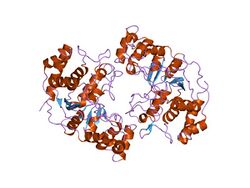
 Crystal structure of flap endonuclease-1 r42e mutant from Pyrococcus horikoshii | |||||||||
| Identifiers | |||||||||
| Symbol | XPG_N | ||||||||
| Pfam | PF00752 | ||||||||
| Pfam clan | CL0280 | ||||||||
| InterPro | IPR006085 | ||||||||
| PROSITE | PDOC00658 | ||||||||
| SCOP2 | 1a77 / SCOPe / SUPFAM | ||||||||
| |||||||||
In molecular biology the protein domain XPG refers to, in this case, the N-terminus of XPG. The XPG protein can be corrected by a 133 kDa nuclear protein, XPGC.[1] XPGC is an acidic protein that confers normal ultraviolet (UV) light resistance.[2] It is a magnesium-dependent, single-strand DNA endonuclease that makes structure-specific endonucleolytic incisions in a DNA substrate containing a duplex region and single-stranded arms.[2][3] XPGC cleaves one strand of the duplex at the border with the single-stranded region.[3]
Homology
XPG belongs to a family of proteins that includes:
- RAD2 from Saccharomyces cerevisiae (Baker's yeast) and rad13 from Schizosaccharomyces pombe (Fission yeast), which are single-stranded DNA endonucleases,;[3][4]
- mouse and human FEN-1, a structure-specific endonuclease;
- RAD2 from fission yeast and RAD27 from budding yeast;
- fission yeast exo1, a 5'-3' double-stranded DNA exonuclease that may act in a pathway that corrects mismatched base pairs;
- yeast DHS1,
- yeast DIN7.
Sequence alignment of this family of proteins reveals that similarities are largely confined to two regions. The first is located at the N-terminal extremity (N-region) and corresponds to the first 95 to 105 amino acids. The second region is internal (I-region) and found towards the C terminus; it spans about 140 residues and contains a highly conserved core of 27 amino acids that includes a conserved pentapeptide (E-A-[DE]-A-[QS]). It is possible that the conserved acidic residues are involved in the catalytic mechanism of DNA excision repair in XPG. The amino acid linking the N- and I-regions are not conserved.
Xeroderma pigmentosum
Xeroderma pigmentosum (XP) [1] is a human autosomal recessive disease, characterised by a high incidence of sunlight-induced skin cancer. People's skin cells with this condition are hypersensitive to ultraviolet light, due to defects in the incision step of DNA excision repair. There are a minimum of seven genetic complementation groups involved in this pathway: XP-A to XP-G. XP-G is one of the most rare and phenotypically heterogeneous of XP, showing anything from slight to extreme dysfunction in DNA excision repair.[2][5]
References
- ↑ 1.0 1.1 "Xeroderma pigmentosum and nucleotide excision repair of DNA". Trends Biochem. Sci. 19 (2): 83–6. February 1994. doi:10.1016/0968-0004(94)90040-X. PMID 8160271.
- ↑ 2.0 2.1 2.2 "Isolation of active recombinant XPG protein, a human DNA repair endonuclease". J. Biol. Chem. 269 (23): 15965–8. June 1994. doi:10.1016/S0021-9258(17)33956-X. PMID 8206890.
- ↑ 3.0 3.1 3.2 "XPG endonuclease makes the 3' incision in human DNA nucleotide excision repair". Nature 371 (6496): 432–5. September 1994. doi:10.1038/371432a0. PMID 8090225. Bibcode: 1994Natur.371..432O.
- ↑ "Yeast excision repair gene RAD2 encodes a single-stranded DNA endonuclease". Nature 366 (6453): 365–8. November 1993. doi:10.1038/366365a0. PMID 8247134. Bibcode: 1993Natur.366..365H.
- ↑ "Evolutionary conservation of excision repair in Schizosaccharomyces pombe: evidence for a family of sequences related to the Saccharomyces cerevisiae RAD2 gene". Nucleic Acids Res. 21 (6): 1345–9. March 1993. doi:10.1093/nar/21.6.1345. PMID 8464724.
 |

