Chemistry:Halovir
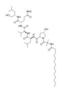
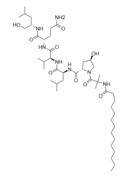
Halovir[1][2][3] refers to a multi-analogue compound belonging to a group of oligopeptides designated as lipopeptaibols (chemical features including lipophilic acyl chain at the N-terminus, abundant α-aminoisobutyric acid content, and a 1,2-amino alcohol located at the C-terminus)[4] which have membrane-modifying capacity[5] and are fungal in origin.[1][6] These peptides display interesting microheterogeneity;[5] slight variation in encoding amino acids gives rise to a mixture of closely related analogues and have been shown to have antibacterial/antiviral properties.[2][6]
Background
Nonribosomal peptides compose a significant group of secondary metabolites in bacterial/fungal organisms (though Drosophila melanogaster and Caenorhabditis elegans both exhibit products of nonribosomal peptide synthetases);[7][8][9] having been observed functioning as self-defense substances/iron-chelating siderophores, they serve as coping mechanisms for environmental stress, perform as virulence factors/toxins promoting pathogenesis, and act in signalling (enabling communications within and between species).[7][8][9] In lieu of these functionalities, many nonribosomal peptides have been utilized in development of medical drugs and biocontrol agents (examples of such include β-lactams, daptomycin, echinocandins, emodepside, bleomycin, cyclosporine, and bialaphos).[7][10][11]
Peptaibols are a family of linear, amphipathic polypeptides (typically consisting of 4-21 amino acids residues)[7] that are generated as a result of the assembly of a variety of aminoacyl, ketoacyl or hydroxyacyl monomers by fungal multimodular megaenzymes denoted as nonribosomal-peptide synthetases (NRPSs).[12] Typically, NRPSs are composed of three highly conserved core domains: an adenylation (A) domain which recognizes, activates and loads monomers onto NRPS, a thiolation (T) domain (also denoted as the peptidyl carrier protein domain) that transports covalently linked monomers/peptidyl intermediates between nearby NRPS domains, and a condensation (C) domain (catalyzes sequential condensation of monomers within the nascent peptide chain).[7][10][13] In addition, a chain-terminating domain [thioesterase (TE) domain, a terminal C (CT) or a reductase (R) domain] is commonly observed at the end of an NRPS in order to relinquish full-length peptide chains in linear or cyclic forms. Furthermore, often seen are feature tailoring domains [epimerase, N-methyltransferase (M), oxidase (Ox), ketoacyl reductase (KR) and cyclase (Cy)], allowing for further modification of monomers/polypeptide intermediates.[7][10][13]
Notable characteristics of peptaibols include: C-terminal alcohol residues (phenylalaninol, leucinol, isoleucinol, valinol),[7][14] an N-acyl terminus (usually acetyl),[14] and high levels of α,α-dialkylated non-proteinogenic amino acids [α-aminoisobutyric acid (Aib), isovaleric acid (Iva), hydroxyproline (Hyp)].[14][15] In most cases, peptaibols form α-helix and β-bend patterns in their 3D structures (α-aminoisobutyric acid is a turn/helix forming agent).[16] α,α-dialkylated amino acid residues in peptaibols create substantial conformation constrictions in the peptide backbone, resulting in the formation of right-handed α-helical structures.[17][18] Membrane modification abilities can be attributed to the formation of transmembrane voltage-dependent channels;[14][19][20] this occurs as the peptide takes on an α-helical conformation upon contact of lipid bilayers, drilling through and forming ion channels with similar electrophysiological configurations of ion channel proteins.[20][21] The principle functionality of the peptides is to rupture membranes, in turn triggering cytolysis via loss of osmotic balance.[21]
Structurally speaking, lipopeptaibols are peptaibols with a fatty acyl moiety linked to the N-terminal amino acid[16] (thusly named), and have been isolated from a number of soil fungi. Their primary structures all have the L-(S-) configuration at the 2-(α-)carbon.[16] They overwhelmingly display microheterogeneity (being very structurally similar; with a limited pool of conserved variation in natural sample).
Structure

[math]\ce{ C45H83N7O9 }[/math]-Halovir A: contains L-leucine, L-valine, and L-glutamine
[math]\ce{ C43H79N7O9 }[/math]-Halovir B: contains L-alanine, L-leucine, L-glutamine
[math]\ce{ C45H83N7O8 }[/math]-Halovir C: contains L-leucine, L-valine, L-glutamine,
[math]\ce{ C43H79N7O9 }[/math]-Halovir D
[math]\ce{ C43H79N7O8 }[/math]-Halovir E
[math]\ce{ C42H77N7O8 }[/math]-Halovir I
[math]\ce{ C44H79N7O9 }[/math]-Halovir J
[math]\ce{ C43H78N7O9 }[/math]-Halovir K
Medical applications
The antibiotic capabilities of these compounds can be attributed to membrane insertion and pore-forming functionalities,[12] and typically exhibit antimicrobial activity in Gram-positive bacteria and fungi.[14][23]
Halovirs A-E (isolated from Scytidium sp.) has displayed potent antiviral activity against HSV-1, and has been observed performing replication inhibition of HSV-1 and HSV-2[24] in standard plaque reduction assay without cytotoxicity (at concentrations upwards of 0.85μM)[24] Mechanistic studies suggest that halovirs kills virus in direct contact and in a time-dependent manner before it can affect host cells.
Halovirs I and J were analyzed for antibacterial and cytotoxic activities, and displayed significant growth inhibition against two Gram-positive bacteria (Staphylococcus aureus and Enterococcus faecium), but not Gram-negative Escherichia coli.[7] Notably, these two halovirs were found to be effective against methicillin-resistant S. aureus (MRSA) and vancomycin-resistant E. faeccium, indicating that the activity against them to be persistent.[7] Additionally, strong cytotoxic activity was observed against a panel of cancer cell lines, including: human lung carinoma A549, human breast carcinoma MCF-7, and human cervical carcinoma HeLa cells (cytotoxicity was not specific to cancer cells in the referenced study).[7]
References
- ↑ 1.0 1.1 "Halovir, an antiviral marine natural product, and derivatives thereof - Patent US-2003013659-A1 - PubChem". https://pubchem.ncbi.nlm.nih.gov/patent/US-2003013659-A1.
- ↑ 2.0 2.1 Rowley, David C.; Kelly, Sara; Kauffman, Christopher A.; Jensen, Paul R.; Fenical, William (2004-02-10). "Halovirs A-E, New Antiviral Agents from a Marine-Derived Fungus of the Genus Scytalidium." (in en). ChemInform 35 (6). doi:10.1002/chin.200406159. ISSN 0931-7597. https://onlinelibrary.wiley.com/doi/10.1002/chin.200406159.
- ↑ Rowley, David C.; Kelly, Sara; Jensen, Paul; Fenical, William (September 2004). "Synthesis and structure–activity relationships of the halovirs, antiviral natural products from a marine-derived fungus" (in en). Bioorganic & Medicinal Chemistry 12 (18): 4929–4936. doi:10.1016/j.bmc.2004.06.044. PMID 15336272. https://linkinghub.elsevier.com/retrieve/pii/S096808960400495X.
- ↑ Toniolo, C.; Crisma, M.; Formaggio, F.; Peggion, C.; Epand, R.F.; Epand, R.M. (August 2001). "Lipopeptaibols, a novel family of membrane active, antimicrobial peptides" (in en). Cellular and Molecular Life Sciences 58 (9): 1179–1188. doi:10.1007/PL00000932. ISSN 1420-682X. PMID 11577977. http://link.springer.com/10.1007/PL00000932.
- ↑ 5.0 5.1 Auvin-Guette, Catherine; Rebuffat, Sylvie; Prigent, Yann; Bodo, Bernard (March 1992). "Trichogin A IV, an 11-residue lipopeptaibol from Trichoderma longibrachiatum" (in en). Journal of the American Chemical Society 114 (6): 2170–2174. doi:10.1021/ja00032a035. ISSN 0002-7863. https://pubs.acs.org/doi/abs/10.1021/ja00032a035.
- ↑ 6.0 6.1 Xiao, Dongliang; Zhang, Mei; Wu, Ping; Li, Tianyi; Li, Wenhua; Zhang, Liwen; Yue, Qun; Chen, Xinqi et al. (May 2022). "Halovirs I–K, antibacterial and cytotoxic lipopeptaibols from the plant pathogenic fungus Paramyrothecium roridum NRRL 2183" (in en). The Journal of Antibiotics 75 (5): 247–257. doi:10.1038/s41429-022-00517-7. ISSN 0021-8820. PMID 35288678. https://www.nature.com/articles/s41429-022-00517-7.
- ↑ 7.0 7.1 7.2 7.3 7.4 7.5 7.6 7.7 7.8 7.9 Xiao, Dongliang; Zhang, Mei; Wu, Ping; Li, Tianyi; Li, Wenhua; Zhang, Liwen; Yue, Qun; Chen, Xinqi et al. (May 2022). "Halovirs I–K, antibacterial and cytotoxic lipopeptaibols from the plant pathogenic fungus Paramyrothecium roridum NRRL 2183" (in en). The Journal of Antibiotics 75 (5): 247–257. doi:10.1038/s41429-022-00517-7. ISSN 0021-8820. PMID 35288678. https://www.nature.com/articles/s41429-022-00517-7.
- ↑ 8.0 8.1 Richardt, Arnd; Kemme, Tobias; Wagner, Stefanie; Schwarzer, Dirk; Marahiel, Mohamed A.; Hovemann, Bernhard T. (2003-10-17). "Ebony, a novel nonribosomal peptide synthetase for beta-alanine conjugation with biogenic amines in Drosophila". The Journal of Biological Chemistry 278 (42): 41160–41166. doi:10.1074/jbc.M304303200. ISSN 0021-9258. PMID 12900414.
- ↑ 9.0 9.1 Shou, Qingyao; Feng, Likui; Long, Yaoling; Han, Jungsoo; Nunnery, Joshawna K.; Powell, David H.; Butcher, Rebecca A. (October 2016). "A hybrid polyketide-nonribosomal peptide in nematodes that promotes larval survival". Nature Chemical Biology 12 (10): 770–772. doi:10.1038/nchembio.2144. ISSN 1552-4469. PMID 27501395.
- ↑ 10.0 10.1 10.2 Süssmuth, Roderich; Müller, Jane; Döhren, Hans von; Molnár, István (2010-12-17). "Fungal cyclooligomer depsipeptides: From classical biochemistry to combinatorial biosynthesis" (in en). Natural Product Reports 28 (1): 99–124. doi:10.1039/C001463J. ISSN 1460-4752. PMID 20959929. https://pubs.rsc.org/en/content/articlelanding/2011/np/c001463j.
- ↑ Felnagle, Elizabeth A.; Jackson, Emily E.; Chan, Yolande A.; Podevels, Angela M.; Berti, Andrew D.; McMahon, Matthew D.; Thomas, Michael G. (2008-04-01). "Nonribosomal Peptide Synthetases Involved in the Production of Medically Relevant Natural Products" (in en). Molecular Pharmaceutics 5 (2): 191–211. doi:10.1021/mp700137g. ISSN 1543-8384. PMID 18217713.
- ↑ 12.0 12.1 Hou, Xuewen; Sun, Ruonan; Feng, Yanyan; Zhang, Runfang; Zhu, Tianjiao; Che, Qian; Zhang, Guojian; Li, Dehai (2022-09-01). "Peptaibols: Diversity, bioactivity, and biosynthesis" (in en). Engineering Microbiology 2 (3): 100026. doi:10.1016/j.engmic.2022.100026. ISSN 2667-3703.
- ↑ 13.0 13.1 Bills, Gerald; Li, Yan; Chen, Li; Yue, Qun; Niu, Xue-Mei; An, Zhiqiang (2014-09-11). "New insights into the echinocandins and other fungal non-ribosomal peptides and peptaibiotics" (in en). Natural Product Reports 31 (10): 1348–1375. doi:10.1039/C4NP00046C. ISSN 1460-4752. PMID 25156669. https://pubs.rsc.org/en/content/articlelanding/2014/np/c4np00046c.
- ↑ 14.0 14.1 14.2 14.3 14.4 Wiest, Aric; Grzegorski, Darlene; Xu, Bi-Wen; Goulard, Christophe; Rebuffat, Sylvie; Ebbole, Daniel J.; Bodo, Bernard; Kenerley, Charles (2002-06-07). "Identification of Peptaibols from Trichoderma virens and Cloning of a Peptaibol Synthetase *" (in English). Journal of Biological Chemistry 277 (23): 20862–20868. doi:10.1074/jbc.M201654200. ISSN 0021-9258. PMID 11909873. https://www.jbc.org/article/S0021-9258(20)84940-0/abstract.
- ↑ Whitmore, Lee; Wallace, B. A. (2004-01-01). "The Peptaibol Database: a database for sequences and structures of naturally occurring peptaibols". Nucleic Acids Research 32 (Database issue): D593–D594. doi:10.1093/nar/gkh077. ISSN 0305-1048. PMID 14681489.
- ↑ 16.0 16.1 16.2 Toniolo, C.; Crisma, M.; Formaggio, F.; Peggion, C.; Epand, R.F.; Epand, R.M. (August 2001). "Lipopeptaibols, a novel family of membrane active, antimicrobial peptides" (in en). Cellular and Molecular Life Sciences 58 (9): 1179–1188. doi:10.1007/PL00000932. ISSN 1420-682X. PMID 11577977. http://link.springer.com/10.1007/PL00000932.
- ↑ Marshall, G R; Hodgkin, E E; Langs, D A; Smith, G D; Zabrocki, J; Leplawy, M T (January 1, 1990). "Factors governing helical preference of peptides containing multiple alpha,alpha-dialkyl amino acids.". Proceedings of the National Academy of Sciences of the United States of America 87 (1): 487–491. doi:10.1073/pnas.87.1.487. ISSN 0027-8424. PMID 2296604. Bibcode: 1990PNAS...87..487M.
- ↑ Schweitzer-Stenner, Reinhard; Gonzales, Widalys; Bourne, Gregory T.; Feng, Jianwen A.; Marshall, Garland R. (2007-10-01). "Conformational Manifold of α-Aminoisobutyric Acid (Aib) Containing Alanine-Based Tripeptides in Aqueous Solution Explored by Vibrational Spectroscopy, Electronic Circular Dichroism Spectroscopy, and Molecular Dynamics Simulations" (in en). Journal of the American Chemical Society 129 (43): 13095–13109. doi:10.1021/ja0738430. ISSN 0002-7863. PMID 17918837. https://pubs.acs.org/doi/10.1021/ja0738430.
- ↑ Rebuffat, S.; Duclohier, H.; Auvin-Guette, C.; Molle, G.; Spach, G.; Bodo, B. (September 1992). "Membrane-modifying properties of the pore-forming peptaibols saturnisporin SA IV and harzianin HA V". FEMS Microbiology Immunology 5 (1–3): 151–160. doi:10.1111/j.1574-6968.1992.tb05886.x. ISSN 0920-8534. PMID 1384595.
- ↑ 20.0 20.1 Sansom, Mark S. P.; Kerr, Ian D.; Mellor, Ian R. (1991-11-01). "Ion channels formed by amphipathic helical peptides" (in en). European Biophysics Journal 20 (4): 229–240. doi:10.1007/BF00183460. ISSN 1432-1017. PMID 1725513. https://doi.org/10.1007/BF00183460.
- ↑ 21.0 21.1 Huang, Huey W. (1996), Merz, Kenneth M.; Roux, Benoît, eds., "Structural Basis and Energetics of Peptide Membrane Interactions" (in en), Biological Membranes: A Molecular Perspective from Computation and Experiment (Boston, MA: Birkhäuser): pp. 281–298, doi:10.1007/978-1-4684-8580-6_9, ISBN 978-1-4684-8580-6, https://doi.org/10.1007/978-1-4684-8580-6_9, retrieved 2022-10-10
- ↑ "Support | pymol.org". https://pymol.org/2/support.html?.
- ↑ Jen, W. -C.; Jones, G. A.; Brewer, D.; Parkinson, V. O.; Taylor, A. (October 1987). "The antibacterial activity of alamethicins and zervamicins" (in en). Journal of Applied Bacteriology 63 (4): 293–298. doi:10.1111/j.1365-2672.1987.tb02705.x. PMID 3436854. https://onlinelibrary.wiley.com/doi/10.1111/j.1365-2672.1987.tb02705.x.
- ↑ 24.0 24.1 Rowley, David C.; Kelly, Sara; Kauffman, Christopher A.; Jensen, Paul R.; Fenical, William (2004-02-10). "Halovirs A-E, New Antiviral Agents from a Marine-Derived Fungus of the Genus Scytalidium." (in en). ChemInform 35 (6). doi:10.1002/chin.200406159. ISSN 0931-7597. https://onlinelibrary.wiley.com/doi/10.1002/chin.200406159.
 |


