Physics:Campanile probe


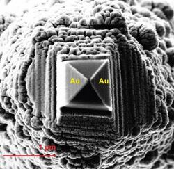
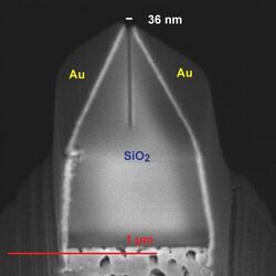
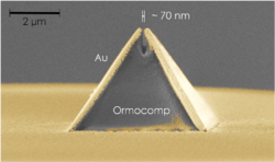
In near-field scanning optical microscopy the campanile probe is a tapered optical probe with a shape of a campanile (a square pyramid). It is made of an optically transparent dielectric, typically silica, and its two facets are coated with a metal, typically gold. At the probe tip, the metal-coated facets are separated by a gap of a few tens of nanometers, which determines the spatial resolution of the probe. Such a probe design allows collecting optical signals, usually photoluminescence (PL) or Raman scattering, with a subwavelength resolution, breaking the diffraction limit.[1][3]
The campanile probe is attached to an optical fiber, which both provides a laser excitation of the studied sample and collects the measured signal. The probe is rastered over the sample with a standard scanning probe microscopy scanner, keeping the distance to the sample surface at a few nanometers.[1] Contrary to the traditional (circular) near-field probes, the campanile probe has no cut-off frequency and is insensitive to the spatial mode of the optical near field. Hence its application is not limited to thin-film samples.[3] Another advantage of the campanile probe is a high signal collection efficiency, which exceeds 90%.[4]
Campanile probes are typically fabricated as follows: a standard cylindrical single-mode optical fiber is etched with hydrofluoric acid to create a conical tip with a radius of ca. 100 nm. Then a square pyramid is carved on the tip using focused ion beam (FIB) milling, and its two facets are coated with a metal by shadow evaporation. A nanometer gap is then opened on the tip by FIB.[3] Alternative fabrication method uses nanoimprint lithography to replicate campanile pyramid from a mold. This approach significantly increases fabrication speed.[2]
References
- ↑ 1.0 1.1 1.2 1.3 1.4 Bao, Wei; Borys, Nicholas J.; Ko, Changhyun; Suh, Joonki; Fan, Wen; Thron, Andrew; Zhang, Yingjie; Buyanin, Alexander et al. (2015). "Visualizing nanoscale excitonic relaxation properties of disordered edges and grain boundaries in monolayer molybdenum disulfide". Nature Communications 6: 7993. doi:10.1038/ncomms8993. PMID 26269394. Bibcode: 2015NatCo...6.7993B.
- ↑ 2.0 2.1 Calafiore, Giuseppe; Koshelev, Alexander; Darlington, Thomas P.; Borys, Nicholas J.; Melli, Mauro; Polyakov, Aleksandr; Cantarella, Giuseppe; Allen, Frances I. et al. (2017-05-10). "Campanile Near-Field Probes Fabricated by Nanoimprint Lithography on the Facet of an Optical Fiber" (in En). Scientific Reports 7 (1): 1651. doi:10.1038/s41598-017-01871-5. ISSN 2045-2322. PMID 28490793. Bibcode: 2017NatSR...7.1651C.
- ↑ 3.0 3.1 3.2 Bao, W.; Melli, M.; Caselli, N.; Riboli, F.; Wiersma, D. S.; Staffaroni, M.; Choo, H.; Ogletree, D. F. et al. (2012). "Mapping Local Charge Recombination Heterogeneity by Multidimensional Nanospectroscopic Imaging". Science 338 (6112): 1317–21. doi:10.1126/science.1227977. PMID 23224550. Bibcode: 2012Sci...338.1317B.
- ↑ Chapelle, Marc Lamy de la; Gucciardi, Pietro Giuseppe; Lidgi-Guigui, Nathalie (28 October 2015). Handbook of Enhanced Spectroscopy. Pan Stanford Publishing. pp. 366–. ISBN 978-981-4613-33-0. https://books.google.com/books?id=fTPSCgAAQBAJ&pg=PA366.
 |
