Chemistry:DiI

| |
| Names | |
|---|---|
| IUPAC name
(2Z)-2-[(E)-3-(3,3-dimethyl-1-octadecylindol-1-ium-2-yl)prop-2-enylidene]-3,3-dimethyl-1-octadecylindole; perchlorate
| |
| Other names
3H-Indolium, 2-[3-(1,3-dihydro-3,3-di-methyl-1-octadecyl-2H-indol-2-ylidene)-1-propenyl]-3,3-dimethyl-1-octadecyl, perchlorate; 1,1'-dioctadecyl-3,3,3'3'-tetramethylindocarbocyanine perchlorate; D 282; DiI; DiIC18(3)
| |
| Identifiers | |
3D model (JSmol)
|
|
| ChemSpider | |
PubChem CID
|
|
| |
| |
| Properties | |
| C59H97ClN2O4 | |
| Molar mass | 933.89 g·mol−1 |
| Melting point | 68 °C (154 °F; 341 K) (decomposes) |
| Solubility | Soluble in ethanol, methanol, DMF, DMSO |
Except where otherwise noted, data are given for materials in their standard state (at 25 °C [77 °F], 100 kPa). | |
| Infobox references | |

DiI, pronounced like Dye Aye, also known as DiIC18(3), is a fluorescent lipophilic cationic indocarbocyanine dye and indolium compound, which is usually made as a perchlorate salt. It is used for scientific staining purposes such as single molecule imaging, fate mapping, electrode marking and neuronal tracing (as DiI is retained in the lipid bilayers).
DiI is manufactured by Invitrogen, which has a series of long-chain lipophilic carbocyanine dyes, of which DiI is one of the most well researched members. Some prominent members of the series includes: DiI, also called DiIC18(3); DiO, also called DiOC18(3); DiD, also called DiIC18(5); and DiR, also called DiIC18(7), which exhibit distinct orange, green, red and infrared fluorescence, respectively, and all have the following useful properties, according to the manufacturer:[1][2]
- Diffuse laterally to stain the entire cell
- Fluoresce weakly in water but highly fluorescent and quite photostable when incorporated into membranes
- Possess very bright signals with high extinction coefficients
- Are well retained in cell membranes
- Demonstrate very little transfer to other cells
Name and Chemical Structure
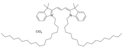
The "Di" possibly stands for the di-alkyl nature and the "I" of DiI possibly stands for the "indocarbocyanine" group (which it shares with D383, D384, D3886, D3899, D3911, D7756, D7776, D7777, D12730, N22880). The full chemical name of this major member of the group is 1,1'-dioctadecyl-3,3,3'3'-tetramethylindocarbocyanine perchlorate, the chemical formula is C59H97ClN2O4 It has 18-carbon-long straight alkyl hydrocarbon tails on each on the nitrogen (Position 1) of the two indoline rings, a conjugated 3-carbon bridge connecting the 2nd positions (Carbon) on the rings symmetrically, and two methyl groups each on each of the 3rd positions (carbon) of the two rings. The longer names e.g. DiIC18(3) mention the length of the alkyl chain (e.g. 18) and the length of the conjugated bridge between the aromatic rings e.g. 3.
Derivatives and Analogs
DiI has several derivatives and analogs in use:[3]
- CM-DiI (C-7000, C-7001) is a fixable analogue of DiI. It has a thiol-reactive chloromethyl (CM) group that makes it more soluble in aqueous solution i.e. culture media and aldehyde-fixable.[4] It has together with SP-DiIC18(3) been demonstrated to be compatible with the tissue clearing method CLARITY, that relies on an extensive lipid removal by detergent (SDS).[5]
- FAST DiI (D-3899 oil, D-7756 solid crystals) has diunsaturated linoleyl (C18:2; Δ9,12) tails in place of the saturated octadecyl tails (C18:0). It reportedly migrates ~50% faster than DiI within membranes.
- Monounsaturated DiI analog (D-3886, oil) with oleate (C18:1, Δ9) tails is also available. It might also migrate faster than DiI within the membrane.
- 5,5′-Ph2-DiIC18(3) (D-7779) has phenyl substituents attached to the indoline rings of DiI, making it more lipophilic and significantly more fluorescent.[6] It has higher quantum yield and longer-wavelength spectra (red-shifted by ~20 nm).
- Anionic DiI analogs with potentially enhanced solubility in culture media:
- Sulfonate analogs e.g.: D-7776 or DiIC18(3)-DS
- Sulfophenyl (SP) analogs e.g.: D-7777 or SP-DiIC18(3)
- Shorter chain DiI analogs:
- D-383 or DiIC12(3)
- D-384 or DiIC16(3)
- DiI and DiO analogs with tails much shorter than 12C, usually less than 7C are used as membrane-potential sensors for mitochondria, based on their vertical sinking into or floating up with respect to a lipid bilayer driven by electric field which changes their fluorescence due to the change in the ambient hydrophobic and charge environment around the fluorescent aromatic rings (they are sensors based on physical movement of whole molecule thus slower compared to membrane potential sensors that work by electron redistribution). Analogs with both intermediate and short tails may be used as organelle stains e.g. of the endoplasmic reticulum.
- Longer wavelength excitable analogs (longer bridges)
- "DiD" or DiIC18(5) (D-307,D-7757) has a 5 carbon bridge, and long-wavelength visible light–excitable analog with red emission
- "DiR" or DiIC18(7) (D-12731) has a 7 carbon bridge, with excitation and emission both in the infrared range thus may not be visible without a CCD or other sensing device that can capture optical signal of infrared nature. But they cause comparative less photodamage to tissue. Thus they can be conveniently used for in vivo imaging.[7]
Physical Properties
The dye (DiIC18(3)) is a violet crystal that is soluble in ethanol, methanol, dimethylformamide, and dimethylsulfoxide.[8] The crystal form of the dye has melting point of 68 °C (at which it decomposes). The dye had an absorption maximum at 549 nm and an emission maximum 565 nm[8] similar to tetramethyl rhodamine. It is mildly fluorescent in aqueous suspension, but becomes bright when bound to cell membrane.[9] Once bound to a membrane it diffuses laterally in 2 dimensions; if there is no diffusion barrier, DiI generally stains the whole leaflet (one surface) of a biological membrane rapidly, but does not readily flip across to the other leaflet.
See also
References
- ↑ Invitrogen Molecular probes Handbook
- ↑ detailed data from invitrogen on this series of lipophilic dyes
- ↑ "Probes Chapter 14 — Fluorescent Tracers of Cell Morphology and Fluid Flow". http://www.mobitec.com/probes/docs/sections/1404.pdf.
- ↑ "Catalog of Cell Biology Products - Carbocyanine Membrane Dyes for Neuronal Tracing". http://probes.invitrogen.com/crawl/lit/catalog/3/sections/0925.html.
- ↑ Jensen, Kristian H. R.; Berg, Rune W. (2016-09-06). "CLARITY-compatible lipophilic dyes for electrode marking and neuronal tracing" (in en). Scientific Reports 6: 32674. doi:10.1038/srep32674. ISSN 2045-2322. PMID 27597115. Bibcode: 2016NatSR...632674J.
- ↑ "molecular probes 2001 product information". http://www.mobitec.com/probes/docs/media/pis/mp00282.pdf.
- ↑ Shan, L. (2004). "Near-infrared fluorescence 1,1-dioctadecyl-3,3,3,3-tetramethylindotricarbocyanine iodide (DiR)-labeled macrophages for cell imaging". Molecular Imaging and Contrast Agent Database (MICAD). National Center for Biotechnology Information (US). https://www.ncbi.nlm.nih.gov/books/NBK23531/.
- ↑ 8.0 8.1 R. W. Sabnis (2010). Handbook of Biological Dyes and Stains: Synthesis and Industrial Applications. John Wiley & Sons. ISBN 9780470586235.
- ↑ "Dil Stain (1,1'-Dioctadecyl-3,3,3',3'-Tetramethylindocarbocyanine Perchlorate ('DiI'; DiIC18(3)))". http://products.invitrogen.com/ivgn/product/D282.
 |

