Biology:SEP15
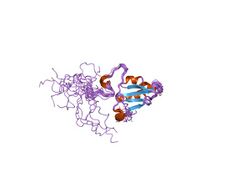
 Generic protein structure example |
15 kDa selenoprotein is a protein that in humans is encoded by the SEP15 gene.[1] Two alternatively spliced transcript variants encoding distinct isoforms have been found for this gene.
Function
This gene encodes a selenoprotein, which contains a selenocysteine (Sec) residue at its active site. The selenocysteine is encoded by the UGA codon that normally signals translation termination. The 3' UTR of selenoprotein genes have a common stem-loop structure, the sec insertion sequence (SECIS), that is necessary for the recognition of UGA as a Sec codon rather than as a stop signal. Studies in mouse suggest that this selenoprotein may have redox function and may be involved in the quality control of protein folding.[1]
Clinical significance
This gene is localized on chromosome 1p31, a genetic locus commonly mutated or deleted in human cancers.[1]
Protein domain
| Sep15 | |||||||||
|---|---|---|---|---|---|---|---|---|---|
 Solution structure of SelM from Mus musculus | |||||||||
| Identifiers | |||||||||
| Symbol | Sep15_SelM | ||||||||
| Pfam | PF08806 | ||||||||
| InterPro | IPR014912 | ||||||||
| |||||||||
The protein this gene encodes for is often called Sep15 however in the case of mice, it is named SelM. This protein is a selenoprotein only found in eukaryotes. This domain has a thioredoxin-like domain and a surface accessible active site redox motif.[2] This suggests that they function as thiol-disulfide isomerases involved in disulfide bond formation in the endoplasmic reticulum.[2]
Function
Recent studies have shown in mice, where the SEP15 gene has been silenced the mice subsequently became deficient in SEP15 and were able to inhibit the development of colorectal cancer.[3]
Structure
The particular structure has an alpha/beta central domain which is actually made up of three alpha helices and a mixed parallel/anti-parallel four-stranded beta-sheet.[2]
References
- ↑ 1.0 1.1 1.2 "Entrez Gene: SEP15 15 kDa selenoprotein". https://www.ncbi.nlm.nih.gov/sites/entrez?Db=gene&Cmd=ShowDetailView&TermToSearch=9403.
- ↑ 2.0 2.1 2.2 "NMR structures of the selenoproteins Sep15 and SelM reveal redox activity of a new thioredoxin-like family". The Journal of Biological Chemistry 281 (6): 3536–43. February 2006. doi:10.1074/jbc.M511386200. PMID 16319061. http://digitalcommons.unl.edu/cgi/viewcontent.cgi?article=1064&context=biochemgladyshev.
- ↑ "Deficiency in the 15 kDa selenoprotein inhibits human colon cancer cell growth". Nutrients 3 (9): 805–17. September 2011. doi:10.3390/nu3090805. PMID 22254125.
Further reading
- "Oligo-capping: a simple method to replace the cap structure of eukaryotic mRNAs with oligoribonucleotides". Gene 138 (1–2): 171–4. January 1994. doi:10.1016/0378-1119(94)90802-8. PMID 8125298.
- "Construction and characterization of a full length-enriched and a 5'-end-enriched cDNA library". Gene 200 (1–2): 149–56. October 1997. doi:10.1016/S0378-1119(97)00411-3. PMID 9373149.
- "A new human selenium-containing protein. Purification, characterization, and cDNA sequence". The Journal of Biological Chemistry 273 (15): 8910–5. April 1998. doi:10.1074/jbc.273.15.8910. PMID 9535873.
- "Structure-expression relationships of the 15-kDa selenoprotein gene. Possible role of the protein in cancer etiology". The Journal of Biological Chemistry 275 (45): 35540–7. November 2000. doi:10.1074/jbc.M004014200. PMID 10945981.
- "Toward a catalog of human genes and proteins: sequencing and analysis of 500 novel complete protein coding human cDNAs". Genome Research 11 (3): 422–35. March 2001. doi:10.1101/gr.GR1547R. PMID 11230166.
- "Association between the 15-kDa selenoprotein and UDP-glucose:glycoprotein glucosyltransferase in the endoplasmic reticulum of mammalian cells". The Journal of Biological Chemistry 276 (18): 15330–6. May 2001. doi:10.1074/jbc.M009861200. PMID 11278576. http://digitalcommons.unl.edu/cgi/viewcontent.cgi?article=1048&context=biochemgladyshev.
- "Genetic and functional analysis of mammalian Sep15 selenoprotein". Protein Sensors and Reactive Oxygen Species - Part A: Selenoproteins and Thioredoxin. Methods in Enzymology. 347. 2002. pp. 187–97. doi:10.1016/S0076-6879(02)47017-6. ISBN 978-0-12-182248-4.
- "[Redox reactions of Sep15 and its relationship with tumor development]". AI Zheng = Aizheng = Chinese Journal of Cancer 22 (2): 119–22. February 2003. PMID 12600282.
- "Exploring proteomes and analyzing protein processing by mass spectrometric identification of sorted N-terminal peptides". Nature Biotechnology 21 (5): 566–9. May 2003. doi:10.1038/nbt810. PMID 12665801.
- "Growth inhibition and induction of apoptosis in mesothelioma cells by selenium and dependence on selenoprotein SEP15 genotype". Oncogene 23 (29): 5032–40. June 2004. doi:10.1038/sj.onc.1207683. PMID 15107826.
- "SMART amplification combined with cDNA size fractionation in order to obtain large full-length clones". BMC Genomics 5 (1): 36. June 2004. doi:10.1186/1471-2164-5-36. PMID 15198809.
 |

