Biology:Ephrin
| Ephrin | |||||||||
|---|---|---|---|---|---|---|---|---|---|
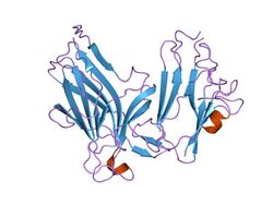
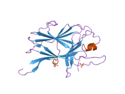
 Ectodomains of the Ephb4-Ephrinb2 protein complex | |||||||||
| Identifiers | |||||||||
| Symbol | Ephrin | ||||||||
| Pfam | PF00812 | ||||||||
| Pfam clan | CL0026 | ||||||||
| InterPro | IPR001799 | ||||||||
| PROSITE | PDOC01003 | ||||||||
| SCOP2 | 1kgy / SCOPe / SUPFAM | ||||||||
| CDD | cd02675 | ||||||||
| Membranome | 70 | ||||||||
| |||||||||
Ephrins (also known as ephrin ligands or Eph family receptor interacting proteins) are a family of proteins that serve as the ligands of the Eph receptor. Eph receptors in turn compose the largest known subfamily of receptor protein-tyrosine kinases (RTKs).
Since ephrin ligands (ephrins) and Eph receptors (Ephs) are both membrane-bound proteins, binding and activation of Eph/ephrin intracellular signaling pathways can only occur via direct cell–cell interaction. Eph/ephrin signaling regulates a variety of biological processes during embryonic development including the guidance of axon growth cones,[1] formation of tissue boundaries,[2] cell migration, and segmentation.[3] Additionally, Eph/ephrin signaling has been identified to play a critical role in the maintenance of several processes during adulthood including long-term potentiation,[4] angiogenesis,[5] and stem cell differentiation.[6]
Classification
Ephrin ligands are divided into two subclasses of ephrin-A and ephrin-B based on their structure and linkage to the cell membrane. Ephrin-As are anchored to the membrane by a glycosylphosphatidylinositol (GPI) linkage and lack a cytoplasmic domain, while ephrin-Bs are attached to the membrane by a single transmembrane domain that contains a short cytoplasmic PDZ-binding motif. The genes that encode the ephrin-A and ephrin-B proteins are designated as EFNA and EFNB respectively. Eph receptors in turn are classified as either EphAs or EphBs based on their binding affinity for either the ephrin-A or ephrin-B ligands.[7]
Of the eight ephrins that have been identified in humans there are five known ephrin-A ligands (ephrin-A1-5) that interact with nine EphAs (EphA1-8 and EphA10) and three ephrin-B ligands (ephrin-B1-3) that interact with five EphBs (EphB1-4 and EphB6).[4][8] Ephs of a particular subclass demonstrate an ability to bind with high affinity to all ephrins of the corresponding subclass, but in general have little to no cross-binding to ephrins of the opposing subclass.[9] However, there are a few exceptions to this intrasubclass binding specificity as it has recently been shown that ephrin-B3 is able to bind to and activate EPH receptor A4 and ephrin-A5 can bind to and activate Eph receptor B2.[10] EphAs/ephrin-As typically bind with high affinity, which can partially be attributed to the fact that ephrinAs interact with EphAs by a "lock-and-key" mechanism that requires little conformational change of the EphAs upon ligand binding. In contrast EphBs typically bind with lower affinity than EphAs/ephring-As since they utilize an "induced fit" mechanism that requires a greater conformational change of EphBs to bind ephrin-Bs.[11]
Function
Axon guidance
During the development of the central nervous system Eph/ephrin signaling plays a critical role in the cell–cell mediated migration of several types of neuronal axons to their target destinations. Eph/ephrin signaling controls the guidance of neuronal axons through their ability to inhibit the survival of axonal growth cones, which repels the migrating axon away from the site of Eph/ephrin activation.[12] The growth cones of migrating axons do not simply respond to absolute levels of Ephs or ephrins in cells that they contact, but rather respond to relative levels of Eph and ephrin expression,[13] which allows migrating axons that express either Ephs or ephrins to be directed along gradients of Eph or ephrin expressing cells towards a destination where axonal growth cone survival is no longer completely inhibited.[12]
Although Eph-ephrin activation is usually associated with decreased growth cone survival and the repulsion of migrating axons, it has recently been demonstrated that growth cone survival does not depend just on Eph-ephrin activation, but rather on the differential effects of "forward" signaling by the Eph receptor or "reverse" signaling by the ephrin ligand on growth cone survival.[12][14]
Retinotopic mapping
The formation of an organized retinotopic map in the superior colliculus (SC) (referred to as the optic tectum in lower vertebrates) requires the proper migration of the axons of retinal ganglion cells (RGCs) from the retina to specific regions in the SC that is mediated by gradients of Eph and ephrin expression in both the SC and in migrating RGCs leaving the retina.[15] The decreased survival of axonal growth cones discussed above allows for a gradient of high posterior to low anterior ephrin-A ligand expression in the SC to direct migrating RGCs axons from the temporal region of the retina that express a high level of EphA receptors toward targets in the anterior SC and RGCs from the nasal retina that have low EphA expression toward their final destination in the posterior SC.[16][17][18] Similarly, a gradient of ephrin-B1 expression along the medial-ventral axis of the SC directs the migration of dorsal and ventral EphB-expressing RGCs to the lateral and medial SC respectively.[19]
Angiogenesis
Ephrins promote angiogenesis in physiological and pathological conditions (e.g. cancer angiogenesis, neovascularisation in cerebral arteriovenous malformation).[20][21] In particular, Ephrin-B2 and EphB4 determine the arterial and venous fate of endothelial cells, respectively, though regulation of angiogenesis by mitigating expression in the VEGF signalling pathway.[20][22] Ephrin-B2 affects VEGF-receptors (e.g.VEGFR3) through forward and reverse signalling pathways.[22] The Ephrin-B2 path extends to lymphangiogenesis, leading to internalization of VEGFR3 in cultured lymphatic endothelial cells.[22] Though the role of ephrins in developmental angiogenesis is elucidated, tumor angiogenesis remains nebulous. Based on observations in Ephrin-A2 deficient mice, Ephrin-A2 may function in forward signalling in tumor angiogenesis; however, this ephrin does not contribute to vascular deformities during development.[23] Moreover, Ephrin-B2 and EphB4 may also contribute to tumor angiogenesis in addition to their positions in development, though the exact mechanism remains unclear.[23] The Ephrin B2/EphB4 and Ephrin B3/EphB1 receptor pairs contribute more to vasculogenesis in addition to angiogenesis whilst Ephrin A1/EphA2 appear to exclusively contribute to angiogenesis.[24]
Several types of Ephrins and Eph receptors have been found to be upregulated in human cancers including breast, colon and liver cancers.[24] Surprisingly, the downregulation of other types of Ephrins and their receptors may also contribute to tumorigenesis; namely, EphA1 in colorectal cancers and EphB6 in melanoma.[24] Displaying similar utility, different ephrins incorporate similar mechanistic pathways to supplement growth of different structures.
Migration factor in intestinal epithelial cell migration
The ephrin protein family of class A and class B guides ligands with the EphB family cell-surface receptors to provide a steady, ordered, and specific migration of the intestinal epithelial cells from the crypt[clarification needed] to villus. The Wnt protein triggers expression of the EphB receptors deep within the crypt, leading to decreased Eph expression and increased ephrin ligand expression, the more superficial a progenitor cell's placement.[25] Migration is caused by a bi-directional signaling mechanism in which the engagement of the ephrin ligand with the EphB receptor regulates the actin cytoskeleton dynamics to cause a "repulsion". Cells remain in place once the interaction ceases to a stop. While the mucus secreting Goblet cells and the absorptive cells move towards the lumen, mature Paneth cells move in the opposite direction, to the bottom of the crypt, where they reside.[26] With the exception of the ephrin ligand binding to EphA5, all other proteins from class A and B have been found in the intestine. However, ephrin proteins A4, A8, B2, and B4 have highest levels in fetal stage, and decline with age.
Experiments performed with Eph receptor knockout mice revealed disorder in the distribution of different cell types.[26] Absorptive cells of various differentiation were mixed with the stem cells within the villi. Without the receptor, the Ephrin ligand was proved to be insufficient for the correct cell placement.[27] Recent studies with knockout mice have also shown evidence of the ephrin-eph interaction indirect role in the suppression of colorectal cancer. The development of adenomatous polyps created by uncontrolled outgrowth of epithelial cells is controlled by ephrin-eph interaction. Mice with APC mutation, without ephrin-B protein lack the means to prevent the spread of ephB positive tumor cells throughout the crypt-villi junction.[28]
Reverse signaling
One unique property of the ephrin ligands is that many have the capacity to initiate a "reverse" signal that is separate and distinct from the intracellular signal activated in Eph receptor-expressing cells. Although the mechanisms by which "reverse" signaling occurs are not completely understood, both ephrin-As and ephrin-Bs have been shown to mediate cellular responses that are distinct from those associated with activation of their corresponding receptors. Specifically, ephrin-A5 was shown to stimulate growth cone spreading in spinal motor neurons[12] and ephrin-B1 was shown to promote dendritic spine maturation.[29]
References
- ↑ "Bidirectional Eph-ephrin signaling during axon guidance". Trends in Cell Biology 17 (5): 230–238. May 2007. doi:10.1016/j.tcb.2007.03.004. PMID 17420126.
- ↑ Hamada, Hiroshi, ed (Mar 2011). "EphrinB/EphB signaling controls embryonic germ layer separation by contact-induced cell detachment". PLOS Biology 9 (3): e1000597. doi:10.1371/journal.pbio.1000597. PMID 21390298.
- ↑ "Ephrin signaling in vivo: look both ways". Developmental Dynamics 232 (1): 1–10. Jan 2005. doi:10.1002/dvdy.20200. PMID 15580616.
- ↑ 4.0 4.1 "Mechanisms and functions of Eph and ephrin signalling". Nature Reviews Molecular Cell Biology 3 (7): 475–486. Jul 2002. doi:10.1038/nrm856. PMID 12094214.
- ↑ "Regulation of angiogenesis by Eph-ephrin interactions". Trends in Cardiovascular Medicine 17 (5): 145–151. Jul 2007. doi:10.1016/j.tcm.2007.03.003. PMID 17574121.
- ↑ "Ephrins and Eph receptors in stem cells and cancer". Current Opinion in Cell Biology 22 (5): 611–616. Oct 2010. doi:10.1016/j.ceb.2010.08.005. PMID 20810264.
- ↑ "Unified nomenclature for Eph family receptors and their ligands, the ephrins. Eph Nomenclature Committee". Cell 90 (3): 403–404. Aug 1997. doi:10.1016/S0092-8674(00)80500-0. PMID 9267020.
- ↑ "Eph/ephrin molecules--a hub for signaling and endocytosis". Genes & Development 24 (22): 2480–2492. Nov 2010. doi:10.1101/gad.1973910. PMID 21078817.
- ↑ "The Eph family of receptors". Current Opinion in Cell Biology 9 (5): 608–615. Oct 1997. doi:10.1016/S0955-0674(97)80113-5. PMID 9330863.
- ↑ "Repelling class discrimination: ephrin-A5 binds to and activates EphB2 receptor signaling". Nature Neuroscience 7 (5): 501–509. May 2004. doi:10.1038/nn1237. PMID 15107857.
- ↑ "Ectodomain structures of Eph receptors". Seminars in Cell & Developmental Biology 23 (1): 35–42. Feb 2012. doi:10.1016/j.semcdb.2011.10.025. PMID 22044883.
- ↑ 12.0 12.1 12.2 12.3 "Coexpressed EphA receptors and ephrin-A ligands mediate opposing actions on growth cone navigation from distinct membrane domains". Cell 121 (1): 127–139. Apr 2005. doi:10.1016/j.cell.2005.01.020. PMID 15820684.
- ↑ "A relative signalling model for the formation of a topographic neural map". Nature 431 (7010): 847–853. Oct 2004. doi:10.1038/nature02957. PMID 15483613. Bibcode: 2004Natur.431..847R.
- ↑ "Ephrin-B2 elicits differential growth cone collapse and axon retraction in retinal ganglion cells from distinct retinal regions". Developmental Neurobiology 70 (11): 781–794. Sep 2010. doi:10.1002/dneu.20821. PMID 20629048.
- ↑ "Eph and ephrin signaling in the formation of topographic maps". Seminars in Cell & Developmental Biology 23 (1): 7–15. Feb 2012. doi:10.1016/j.semcdb.2011.10.026. PMID 22044886.
- ↑ "Multiple roles of EPH receptors and ephrins in neural development". Nature Reviews. Neuroscience 2 (3): 155–164. Mar 2001. doi:10.1038/35058515. PMID 11256076.
- ↑ "Complementary gradients in expression and binding of ELF-1 and Mek4 in development of the topographic retinotectal projection map". Cell 82 (3): 371–381. Aug 1995. doi:10.1016/0092-8674(95)90426-3. PMID 7634327.
- ↑ "In vitro guidance of retinal ganglion cell axons by RAGS, a 25 kDa tectal protein related to ligands for Eph receptor tyrosine kinases". Cell 82 (3): 359–370. Aug 1995. doi:10.1016/0092-8674(95)90425-5. PMID 7634326.
- ↑ "Topographic mapping in dorsoventral axis of the Xenopus retinotectal system depends on signaling through ephrin-B ligands". Neuron 35 (3): 461–473. Aug 2002. doi:10.1016/S0896-6273(02)00786-9. PMID 12165469.
- ↑ 20.0 20.1 "Essential roles of EphB receptors and EphrinB ligands in endothelial cell function and angiogenesis". Advances in Cancer Research 114 (2): 21–57. 2012. doi:10.1016/B978-0-12-386503-8.00002-8. ISBN 9780123865038. PMID 22588055.
- ↑ "Ephrin B2 and EphB4 selectively mark arterial and venous vessels in cerebral arteriovenous malformation". The Journal of International Medical Research 42 (2): 405–15. Apr 2014. doi:10.1177/0300060513478091. PMID 24517927.
- ↑ 22.0 22.1 22.2 "Ephrin-B2 controls VEGF-induced angiogenesis and lymphangiogenesis". Nature 465 (7297): 483–486. May 2010. doi:10.1038/nature09002. PMID 20445537. Bibcode: 2010Natur.465..483W.
- ↑ 23.0 23.1 "Eph receptors and ephrins in cancer: bidirectional signalling and beyond". Nature Reviews. Cancer 10 (3): 165–80. Mar 2010. doi:10.1038/nrc2806. PMID 20179713.
- ↑ 24.0 24.1 24.2 Mosch, Birgit; Reissenweber, Bettina; Neuber, Christin; Pietzsch, Jens (2010). "Eph Receptors and Ephrin Ligands: Important Players in Angiogenesis and Tumor Angiogenesis". Journal of Oncology 2010: 1–12. doi:10.1155/2010/135285. ISSN 1687-8450. PMID 20224755.
- ↑ Alberts, Bruce; Johnson, Alexander; lewis, Julian; Raff, Martin; Roberts, Keith; Walter, Peter (2007). Molecular Biology of the Cell. Garland Sciences. p. 1 440–1441. ISBN 978-0815341055. https://archive.org/details/molecularbiology00albe_292.
- ↑ 26.0 26.1 Batlle, Eduard. "Wnt signalling and EphB-ephrin interactions in intestinal stem cells and CRC progression". 2007 Scientific Report. http://www.irbbarcelona.org/files/File/023-wnts-07.pdf.
- ↑ "Developmental expression of Eph and ephrin family genes in mammalian small intestine". Digestive Diseases and Sciences 55 (9): 2478–88. Sep 2010. doi:10.1007/s10620-009-1102-z. PMID 20112066.
- ↑ Pitulescu, Mara (2010). "Eph/ephrin molecules-a hub for signaling and endocytosis". Genes & Development 24 (22): 2480–2492. doi:10.1101/gad.1973910. PMID 21078817.
- ↑ "Grb4 and GIT1 transduce ephrinB reverse signals modulating spine morphogenesis and synapse formation". Nature Neuroscience 10 (3): 301–310. Mar 2007. doi:10.1038/nn1858. PMID 17310244.
 |


