Biology:Mammillary body
| Mammillary body | |
|---|---|
 Sagittal section, "corpus mamillare" highlighted.[1] | |
 Coronal section of brain through intermediate mass of third ventricle. (Label "corpora mamillaria" at bottom.) | |
| Details | |
| Part of | Diencephalon |
| System | Limbic |
| Parts | medial mammillary nucleus lateral mammillary nucleus |
| Identifiers | |
| Latin | Corpus mamillare (Plural: Corpora mamillaria) |
| Acronym(s) | mmb |
| Anatomical terms of neuroanatomy | |
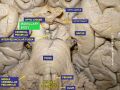
The mammillary bodies are a pair of small round bodies, located on the undersurface of the brain that, as part of the diencephalon, form part of the limbic system. They are located at the ends of the anterior arches of the fornix.[2] They consist of two groups of nuclei, the medial mammillary nuclei and the lateral mammillary nuclei.[3]
Neuroanatomists have often categorized the mammillary bodies as part of the posterior part of hypothalamus.[4]
Structure
Connections
They are connected to other parts of the brain (as shown in the schematic, below left), and act as a relay for impulses coming from the amygdalae and hippocampi, via the mamillo-thalamic tract to the thalamus.
Function
Mammillary body

Mammillary bodies, and their projections to the anterior thalamus via the mammillothalamic tract, are important for recollective memory.[5] The damage of medial mammillary nucleus leads to spatial memory deficit, according to observations in rats with mammillary body lesions.[5]
Clinical significance
Damage to the mammillary bodies due to thiamine deficiency is implied in pathogenesis of Wernicke–Korsakoff syndrome. Symptoms include impaired memory, also called anterograde amnesia, suggesting that the mammillary bodies may be important for memory. Lesions of the medial dorsal and anterior nuclei of the thalami and lesions of the mammillary bodies are commonly involved in amnesic syndromes in humans.[6]
Mammillary body atrophy is present in several other conditions, such as colloid cysts in the third ventricle, Alzheimer’s disease, schizophrenia, heart failure, and sleep apnea. In spite of this the exact function of the mammillary bodies is still not clear.[5]
See also
- Mammillotegmental fasciculus
References
- ↑ Henry Gray (1918). Anatomy of the Human Body.
- ↑ "Mammillary Bodies". Springer Reference. http://www.springerreference.com/docs/html/chapterdbid/183873.html.
- ↑ "The mammillary bodies: two memory systems in one?". Nature Reviews. Neuroscience 5 (1): 35–44. January 2004. doi:10.1038/nrn1299. PMID 14708002. http://www.cf.ac.uk/psych/resources/vann2004.pdf.[yes|permanent dead link|dead link}}].
- ↑ M.B. Carpenter and J. Sutin: Human Neuroanatomy (8th edition) 1983
- ↑ 5.0 5.1 5.2 "Re-evaluating the role of the mammillary bodies in memory". Neuropsychologia 48 (8): 2316–27. July 2010. doi:10.1016/j.neuropsychologia.2009.10.019. PMID 19879886.
- ↑ "Absence of memory dysfunction after bilateral mammillary body and mammillothalamic tract electrode implantation: preliminary experience in three patients". AJNR. American Journal of Neuroradiology 26 (1): 195–7; author reply 197–8. January 2005. PMID 15661728. PMC 7975039. http://www.ajnr.org/cgi/content/full/26/1/195.
External links
- "Anatomy diagram: 13048.000-3". Roche Lexicon - illustrated navigator. Elsevier. http://www.tk.de/rochelexikon/pics/s13048.000-3.html.
 |  |

