Biology:Vitronectin
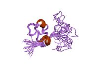
 Generic protein structure example |
Vitronectin (VTN or VN) is a glycoprotein of the hemopexin family which is synthesized and excreted by the liver, and abundantly found in serum, the extracellular matrix and bone.[1] In humans it is encoded by the VTN gene.[2][3]
Vitronectin binds to integrin alpha-V beta-3 and thus promotes cell adhesion and spreading. It also inhibits the membrane-damaging effect of the terminal cytolytic complement pathway and binds to several serpins (serine protease inhibitors). It is a secreted protein and exists in either a single chain form or a clipped, two chain form held together by a disulfide bond.[2] Vitronectin has been speculated to be involved in hemostasis[4] and tumor malignancy.[5][6]
Structure
Vitronectin is a 54 kDa glycoprotein, consisting of 478 amino acid residues. About one-third of the protein's molecular mass is composed of carbohydrates. On occasion, the protein is cleaved after arginine 379, to produce two-chain vitronectin, where the two parts are linked by a disulfide bond. No high-resolution structure has been determined experimentally yet, except for the N-terminal domain.
The protein consists of three domains:
- The N-terminal Somatomedin B domain (1-39)
- A central domains with hemopexin homology (131-342)
- A C-terminal domain (residues 347-459) also with hemopexin homology.
Several structures has been reported for the Somatomedin B domain. The protein was initially crystallized in complex with one of its physiological binding partners: the Plasminogen activator inhibitor-1 (PAI-1) and the structure solved for this complex.[7] Subsequently two groups reported NMR structures of the domain.[8][9]
The somatomedin B domain is a close-knit disulfide knot, with 4 disulfide bonds within 35 residues. Different disulfide configurations had been reported for this domain[10][11][12] but this ambiguity has been resolved by the crystal structure.[12]
Homology models have been built for the central and C-terminal domains.[12]
Function
The somatomedin B domain of vitronectin binds to plasminogen activator inhibitor-1 (PAI-1), and stabilizes it.[7] Thus vitronectin serves to regulate proteolysis initiated by plasminogen activation. In addition, vitronectin is a component of platelets and is, thus, involved in hemostasis. Vitronectin contains an RGD (45-47) sequence, which is a binding site for membrane-bound integrins, e.g., the vitronectin receptor, which serve to anchor cells to the extracellular matrix. The Somatomedin B domain interacts with the urokinase receptor, and this interaction has been implicated in cell migration and signal transduction. High plasma levels of both PAI-1 and the urokinase receptor have been shown to correlate with a negative prognosis for cancer patients. Cell adhesion and migration are directly involved in cancer metastasis, which provides a probable mechanistic explanation for this observation.
References
- ↑ Boron, Walter F. and Boulpaep, Emile L. "Medical Physiology". Saunders, 2012, p.1097.
- ↑ 2.0 2.1 "Entrez Gene: M Vitronectin". https://www.ncbi.nlm.nih.gov/sites/entrez?Db=gene&Cmd=ShowDetailView&TermToSearch=7448.
- ↑ "Nucleotide sequence and organization of the human S-protein gene: repeating peptide motifs in the "pexin" family and a model for their evolution". Biochemistry 26 (21): 6735–42. Oct 1987. doi:10.1021/bi00395a024. PMID 2447940.
- ↑ "Role of vitronectin and its receptors in haemostasis and vascular remodeling". Thrombosis Research 89 (1): 1–21. Jan 1998. doi:10.1016/S0049-3848(97)00298-3. PMID 9610756.
- ↑ "Vitronectin and its receptors". Current Opinion in Cell Biology 5 (5): 864–8. Oct 1993. doi:10.1016/0955-0674(93)90036-P. PMID 7694604.
- ↑ Hurt, Elaine M.; Chan, King; Serrat, Maria Ana Duhagon; Thomas, Suneetha B.; Veenstra, Timothy D.; Farrar, William L. (2009). "Identification of Vitronectin as an Extrinsic Inducer of Cancer Stem Cell Differentiation and Tumor Formation". Stem Cells 28 (3): 390–8. doi:10.1002/stem.271. PMID 19998373.
- ↑ 7.0 7.1 "How vitronectin binds PAI-1 to modulate fibrinolysis and cell migration". Nature Structural Biology 10 (7): 541–4. Jul 2003. doi:10.1038/nsb943. PMID 12808446.
- ↑ "Disulfide bonding arrangements in active forms of the somatomedin B domain of human vitronectin". Biochemistry 43 (21): 6519–34. Jun 2004. doi:10.1021/bi049647c. PMID 15157085.
- ↑ "The solution structure of the N-terminal domain of human vitronectin: proximal sites that regulate fibrinolysis and cell migration". The Journal of Biological Chemistry 279 (28): 29359–66. Jul 2004. doi:10.1074/jbc.M401279200. PMID 15123712.
- ↑ "Identification of the disulfide bonds in the recombinant somatomedin B domain of human vitronectin". The Journal of Biological Chemistry 277 (30): 27109–19. Jul 2002. doi:10.1074/jbc.M200354200. PMID 12019263.
- ↑ "Assignment of the four disulfides in the N-terminal somatomedin B domain of native vitronectin isolated from human plasma". The Journal of Biological Chemistry 279 (34): 35867–78. Aug 2004. doi:10.1074/jbc.M405716200. PMID 15173163.
- ↑ 12.0 12.1 12.2 "Model for the three-dimensional structure of vitronectin: predictions for the multi-domain protein from threading and docking". Proteins 44 (3): 312–20. Aug 2001. doi:10.1002/prot.1096. PMID 11455604.
Further reading
- "Vitronectin in bacterial pathogenesis: a host protein used in complement escape and cellular invasion". Mol. Microbiol. 78 (3): 545–60. 2010. doi:10.1111/j.1365-2958.2010.07373.x. PMID 20807208.
- "Haemophilus influenzae protein E recognizes the C-terminal domain of vitronectin and modulates the membrane attack complex". Mol. Microbiol. 81 (1): 80–98. 2011. doi:10.1111/j.1365-2958.2011.07678.x. PMID 21542857.
- "Haemophilus influenzae acquires vitronectin via the ubiquitous Protein F to subvert host innate immunity". Mol. Microbiol. 87 (6): 1245–66. 2013. doi:10.1111/mmi.12164. PMID 23387957.
External links
- Vitronectin at the US National Library of Medicine Medical Subject Headings (MeSH)
 |


