Biology:Myosin head
| Myosin_head | |||||||||
|---|---|---|---|---|---|---|---|---|---|
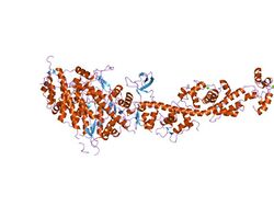
 Scallop myosin in the near-rigor conformation | |||||||||
| Identifiers | |||||||||
| Symbol | Myosin_head | ||||||||
| Pfam | PF00063 | ||||||||
| Pfam clan | CL0023 | ||||||||
| InterPro | IPR001609 | ||||||||
| PROSITE | PDOC00017 | ||||||||
| SCOP2 | 1mys / SCOPe / SUPFAM | ||||||||
| CDD | cd00124 | ||||||||
| |||||||||
The myosin head is the part of the thick myofilament made up of myosin that acts in muscle contraction, by sliding over thin myofilaments of actin. Myosin is the major component of the thick filaments and most myosin molecules are composed of a head, neck, and tail domain; the myosin head binds to thin filamentous actin, and uses ATP hydrolysis to generate force and "walk" along the thin filament. Myosin exists as a hexamer of two heavy chains,[1] two alkali light chains, and two regulatory light chains. The heavy chain can be subdivided into the globular head at the N-terminal and the coiled-coil rod-like tail at the C-terminal, although some forms have a globular region in their C-terminal.
There are many cell-specific isoforms of myosin heavy chains, coded for by a multi-gene family.[2] Myosin interacts with actin to convert chemical energy, in the form of ATP, to mechanical energy.[3] The 3-D structure of the head portion of myosin has been determined [4] and a model for actin-myosin complex has been constructed.[5]
The globular head is well conserved,[4][6][7] and is key to contraction. Muscle contraction results from an attachment–detachment cycle between the myosin heads extending from myosin filaments and the sites on actin filaments. The myosin head first attaches to actin together with the products of ATP hydrolysis, performs a power stroke associated with release of hydrolysis products, and detaches from actin upon binding with new ATP. The detached myosin head then hydrolyses ATP, and performs a recovery stroke to restore its initial position. The strokes have been suggested to result from rotation of the lever arm domain around the converter domain, while the catalytic domain remains rigid.[8]
References
- ↑ "The primary structure of skeletal muscle myosin heavy chain: I. Sequence of the amino-terminal 23 kDa fragment". J. Biochem. 110 (1): 54–9. July 1991. doi:10.1093/oxfordjournals.jbchem.a123543. PMID 1939027.
- ↑ "Human embryonic myosin heavy chain cDNA. Interspecies sequence conservation of the myosin rod, chromosomal locus and isoform specific transcription of the gene". FEBS Lett. 256 (1–2): 21–8. October 1989. doi:10.1016/0014-5793(89)81710-7. PMID 2806546.
- ↑ "Conserved protein domains in a myosin heavy chain gene from Dictyostelium discoideum". Proc. Natl. Acad. Sci. U.S.A. 83 (24): 9433–7. December 1986. doi:10.1073/pnas.83.24.9433. PMID 3540939. Bibcode: 1986PNAS...83.9433W.
- ↑ 4.0 4.1 "Three-dimensional structure of myosin subfragment-1: a molecular motor". Science 261 (5117): 50–8. July 1993. doi:10.1126/science.8316857. PMID 8316857. Bibcode: 1993Sci...261...50R.
- ↑ "Structure of the actin-myosin complex and its implications for muscle contraction". Science 261 (5117): 58–65. July 1993. doi:10.1126/science.8316858. PMID 8316858. Bibcode: 1993Sci...261...58R.
- ↑ "Movement and force produced by a single myosin head". Nature 378 (6553): 209–12. November 1995. doi:10.1038/378209a0. PMID 7477328. Bibcode: 1995Natur.378..209M.
- ↑ "Single-molecule measurement of the stiffness of the rigor myosin head". Biophysical Journal 94 (6): 2160–9. March 2008. doi:10.1529/biophysj.107.119396. PMID 18065470. Bibcode: 2008BpJ....94.2160L.
- ↑ "Electron microscopic evidence for the myosin head lever arm mechanism in hydrated myosin filaments using the gas environmental chamber". Biochemical and Biophysical Research Communications 405 (4): 651–6. February 2011. doi:10.1016/j.bbrc.2011.01.087. PMID 21281603.
 |

