Medicine:Pneumatosis
| Pneumatosis | |
|---|---|
 | |
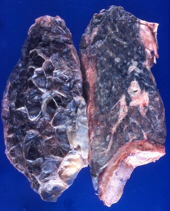
| Left lung completely affected by bullae shown in contrast to a normal lung on the right. | |
| Causes | Tobacco smoking, pollutants |
Pneumatosis is the abnormal presence of air or other gas within tissues.[1]
In the lungs, emphysema involves enlargement of the distal airspaces,[2] and is a major feature of chronic obstructive pulmonary disease (COPD). Other pneumatoses in the lungs are focal (localized) blebs and bullae, pulmonary cysts and cavities.
Pneumoperitoneum (or peritoneal emphysema) is air or gas in the abdominal cavity, and is most commonly caused by gastrointestinal perforation, often the result of surgery.
Pneumarthrosis, the presence of air in a joint, is rarely a serious sign.
Lung cysts

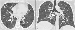
A lung cyst, or pulmonary cyst, encloses a small volume of air, and has a wall thickness of up to 4 mm.[3] A minimum wall thickness of 1 mm has been suggested,[3] but thin-walled pockets may be included in the definition as well.[4] Pulmonary cysts are not associated with either smoking or emphysema.[5]
A lung cavity has a wall thickness of more than 4 mm.[3]
Other thoracic
- Pneumothorax, air or gas in the pleural space
- Pneumomediastinum, air or gas in the mediastinum
- Also called mediastinal emphysema or pneumatosis/emphysema of the mediastinum
Abdominal

- Pneumoperitoneum (or peritoneal emphysema), air or gas in the abdominal cavity. The most common cause is a perforated abdominal viscus, generally a perforated peptic ulcer, although any part of the bowel may perforate from a benign ulcer, tumor or abdominal trauma.
- Pneumatosis intestinalis, air or gas cysts in the bowel wall
- Gastric pneumatosis (or gastric emphysema) is air or gas cysts in the stomach wall[6]
Joints
Pneumarthrosis is the presence of air in a joint. Its presentation on radiography is a radiolucent cleft often called a vacuum phenomenon, or vacuum sign.[7] Pneumarthrosis is associated with osteoarthritis and spondylosis.[8]
Pneumarthrosis is a common normal finding in shoulders[7] as well as in sternoclavicular joints.[9] It is believed to be a cause of the sounds of joint cracking.[8] It is also a common normal post-operative finding at least after spinal surgery.[10] Pneumarthrosis is extremely rare in conjunction with fluid or pus in a joint, and its presence can therefore practically exclude infection.[8]
-
X-ray of a hip with hip replacement and pneumarthrosis, in this case aseptic.
-
A vacuum sign, or vacuum phenomenon, is a normal finding on shoulder X-rays.
Other

Subcutaneous emphysema is found in the deepest layer of the skin. Emphysematous cystitis is a condition of gas in the bladder wall. On occasion this may give rise to secondary subcutaneous emphysema which has a poor prognosis.[11]
Pneumoparotitis is the presence of air in the parotid gland caused by raised air pressure in the mouth often as a result of playing wind instruments. In rare cases air may escape from the gland and give rise to subcutaneous emphysema in the face, neck, or mediastinum.[12][13]
Terminology
The term pneumatosis has word roots of pneumat- + -osis, meaning "air problem/injury".
References
- ↑ "Medical Definition of PNEUMATOSIS" (in en). https://www.merriam-webster.com/medical/pneumatosis.
- ↑ page 64 in: Adrian Shifren (2006). The Washington Manual Pulmonary Medicine Subspecialty Consult, Washington manual subspecialty consult series. Lippincott Williams & Wilkins. ISBN 9780781743761.
- ↑ 3.0 3.1 3.2 Dr Daniel J Bell and Dr Yuranga Weerakkody. "Pulmonary cyst". https://radiopaedia.org/articles/pulmonary-cyst.
- ↑ Araki, Tetsuro; Nishino, Mizuki; Gao, Wei; Dupuis, Josée; Putman, Rachel K; Washko, George R; Hunninghake, Gary M; O'Connor, George T et al. (2015). "Pulmonary cysts identified on chest CT: are they part of aging change or of clinical significance?". Thorax 70 (12): 1156–1162. doi:10.1136/thoraxjnl-2015-207653. ISSN 0040-6376. PMID 26514407.
- ↑ Araki, Tetsuo. "Pulmonary cysts identified on chest CT:are they part of ageing change or of clinical significance". https://thorax.bmj.com/content/thoraxjnl/early/2015/10/29/thoraxjnl-2015-207653.full.pdf.
- ↑ "Gastric emphysema | Radiology Reference Article | Radiopaedia.org". https://radiopaedia.org/articles/gastric-emphysema-1.
- ↑ 7.0 7.1 Abhijit Datir. "Vacuum phenomenon in shoulder". https://radiopaedia.org/articles/vacuum-phenomenon-in-shoulder.
- ↑ 8.0 8.1 8.2 Page 60 in: Harry Griffiths (2008). Musculoskeletal Radiology. CRC Press. ISBN 9781420020663.
- ↑ Restrepo, Carlos S.; Martinez, Santiago; Lemos, Diego F.; Washington, Lacey; McAdams, H. Page; Vargas, Daniel; Lemos, Julio A.; Carrillo, Jorge A. et al. (2009). "Imaging Appearances of the Sternum and Sternoclavicular Joints". RadioGraphics 29 (3): 839–859. doi:10.1148/rg.293055136. ISSN 0271-5333. PMID 19448119.
- ↑ Mall, J C; Kaiser, J A (1987). "The usual appearance of the postoperative lumbar spine.". RadioGraphics 7 (2): 245–269. doi:10.1148/radiographics.7.2.3448634. ISSN 0271-5333. PMID 3448634.
- ↑ "Emphysematous cystitis with clinical subcutaneous emphysema". International Journal of Emergency Medicine 4 (1): 26. June 2011. doi:10.1186/1865-1380-4-26. PMID 21668949.
- ↑ McCormick, Michael E.; Bawa, Gurneet; Shah, Rahul K. (2013). "Idiopathic recurrent pneumoparotitis". American Journal of Otolaryngology 34 (2): 180–182. doi:10.1016/j.amjoto.2012.11.005. ISSN 0196-0709. PMID 23318047.
- ↑ Joiner MC; van der Kogel A (15 June 2016). Basic Clinical Radiobiology, Fifth Edition. CRC Press. p. 1908. ISBN 978-0-340-80893-1. https://books.google.com/books?id=Xp4YtqM7Ef8C&pg=PA1908.
External links
| Classification |
|---|
 |


