Biology:MYND zinc finger
| zf-MYND | |||||||||
|---|---|---|---|---|---|---|---|---|---|
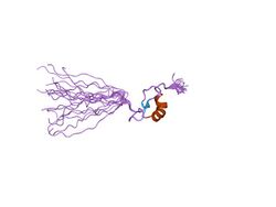
 solution structure of zf-mynd domain of protein cbfa2ti (protein mtg8) | |||||||||
| Identifiers | |||||||||
| Symbol | zf-MYND | ||||||||
| Pfam | PF01753 | ||||||||
| Pfam clan | CL0175 | ||||||||
| InterPro | IPR002893 | ||||||||
| |||||||||
In molecular biology the MYND-type zinc finger domain is a conserved protein domain. The MYND domain (myeloid, Nervy, and DEAF-1) is present in a large group of proteins that includes RP-8 (PDCD2), Nervy, and predicted proteins from Drosophila, mammals, Caenorhabditis elegans, yeast, and plants.[1][2][3] The MYND domain consists of a cluster of cysteine and histidine residues, arranged with an invariant spacing to form a potential zinc-binding motif.[2] Mutating conserved cysteine residues in the DEAF-1 MYND domain does not abolish DNA binding, which suggests that the MYND domain might be involved in protein-protein interactions.[2] Indeed, the MYND domain of ETO/MTG8 interacts directly with the N-CoR and SMRT co-repressors.[4][5] Aberrant recruitment of co-repressor complexes and inappropriate transcriptional repression is believed to be a general mechanism of leukemogenesis caused by the t(8;21) translocations that fuse ETO with the acute myelogenous leukemia 1 (AML1) protein. ETO has been shown to be a co-repressor recruited by the promyelocytic leukemia zinc finger (PLZF) protein.[6] A divergent MYND domain present in the adenovirus E1A binding protein BS69 was also shown to interact with N-CoR and mediate transcriptional repression.[7] The current evidence suggests that the MYND motif in mammalian proteins constitutes a protein-protein interaction domain that functions as a co-repressor-recruiting interface.
References
- ↑ "Identification of homeotic target genes in Drosophila melanogaster including nervy, a proto-oncogene homologue". Genetics 140 (2): 573–86. June 1995. doi:10.1093/genetics/140.2.573. PMID 7498738.
- ↑ 2.0 2.1 2.2 "DEAF-1, a novel protein that binds an essential region in a Deformed response element". EMBO J. 15 (8): 1961–70. April 1996. doi:10.1002/j.1460-2075.1996.tb00547.x. PMID 8617243.
- ↑ "Identification of mRNAs associated with programmed cell death in immature thymocytes". Mol. Cell. Biol. 11 (8): 4177–88. August 1991. doi:10.1128/MCB.11.8.4177. PMID 2072913.
- ↑ "The MYND motif is required for repression of basal transcription from the multidrug resistance 1 promoter by the t(8;21) fusion protein". Mol. Cell. Biol. 18 (6): 3604–11. June 1998. doi:10.1128/MCB.18.6.3604. PMID 9584201.
- ↑ "ETO, a target of t(8;21) in acute leukemia, interacts with the N-CoR and mSin3 corepressors". Mol. Cell. Biol. 18 (12): 7176–84. December 1998. doi:10.1128/MCB.18.12.7176. PMID 9819404.
- ↑ "The ETO protein disrupted in t(8;21)-associated acute myeloid leukemia is a corepressor for the promyelocytic leukemia zinc finger protein". Mol. Cell. Biol. 20 (6): 2075–86. March 2000. doi:10.1128/MCB.20.6.2075-2086.2000. PMID 10688654.
- ↑ "The adenovirus E1A binding protein BS69 is a corepressor of transcription through recruitment of N-CoR". Oncogene 19 (12): 1538–46. March 2000. doi:10.1038/sj.onc.1203421. PMID 10734313.
External links
- Eukaryotic Linear Motif resource motif class LIG_MYND
 |

