Chemistry:Field stain
From HandWiki

Micrograph of a Field stain showing malignant melanoma.
Field stain is a histological method for staining of blood smears. It is used for staining thick blood films in order to discover malarial parasites. Field's stain is a version of a Romanowsky stain, used for rapid processing of the specimens.[1]
Field's stain consists of two parts - Field's stain A is methylene blue and Azure 1 dissolved in phosphate buffer solution; Field's stain B is Eosin Y in buffer solution. Field stain is named after physician John William Field, who developed it in 1941.[2]
Additional images
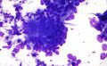
Colorectal adenocarcinoma. Field stain.
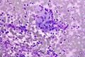
Granuloma. Field stain.


References
- ↑ Chunge, CN.; Ngige, S.; Bwibo, CR.; Mulega, PC.; Kilonzo, JF.; Kibati, F.; Owate, J. (Aug 1989). "A rapid staining technique for Leishmania parasites in splenic aspirate smears". Ann Trop Med Parasitol 83 (4): 361–4. doi:10.1080/00034983.1989.11812358. PMID 2481429.
- ↑ "Obituaries". Medical Journal of Malaysia. June 1981. http://www.e-mjm.org/1981/v36n2/Obituaries.pdf.
External links
 |  |

